Image Size: [small] [small] [as-submitted] [as-submitted]

 |
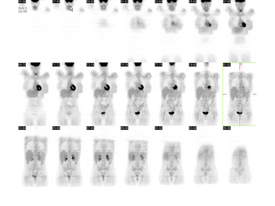
| Fig. 1 |
| Coronal PET images reveal no evidence of increased FDG metabolism to suggest active metastatic disease. |
|

 |
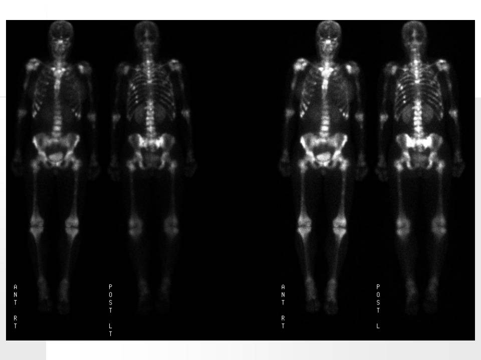
| Fig. 2 |
| Bone scan images reveal innumerable foci of increased activity throughout the mid thoracic and lower lumbar spine and proximal appendicular skeleton. |
|

 |
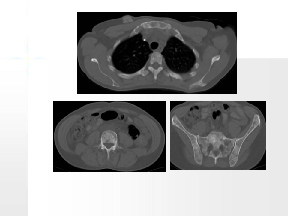
| Fig. 3 |
| CT axial images reveal osteoblastic metastatic disease of the axial skeleton, some of the ribs, the sternum, the scapula, and the pelvis osteoblastic metastatic disease of the axial skeleton, some of the ribs, the sternum, the scapula, and the pelvis.
|
|
Image Size: [small] [small] [as-submitted] [as-submitted]
|