back to index of exported cases

Unknown Case
Authored By: Xiaoni Hong and Jerold Wallis, Assoc Prof of Radiology.
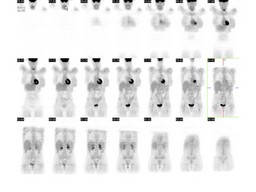
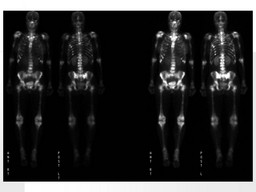
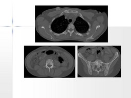
Patient: 40 year old female
History: 40-year-old woman with stage IV left breast cancer diagnosed in October  2005, status post radiation therapy completed in April, 2006, status post chemotherapy completed in June, 2007. Currently, the patient is on hormone therapy with Arimidex.
Image Size:[small][as-submitted]

Fig. 1
Coronal PET images reveal no evidence of increased FDG metabolism to suggest active metastatic disease.

Fig. 2
Bone scan images reveal innumerable foci of increased activity throughout the mid thoracic and lower lumbar spine and proximal appendicular skeleton.

Fig. 3
CT axial images reveal osteoblastic metastatic disease of the axial skeleton, some of the ribs, the sternum, the scapula, and the pelvis osteoblastic metastatic disease of the axial skeleton, some of the ribs, the sternum, the scapula, and the pelvis.
Image Size:[small][as-submitted]

Comments:
No comments posted.
Additional Details:

Case [View Case with Diagnosis]Owner(s): Xiaoni Hong and Jerold Wallis, Assoc Prof of RadiologyLast Updated: 02-07-2013
Anatomy: Skeletal System   Pathology: Other
Modality: Nuc MedAccess Level: Readable by all users, writable by NucMed Certifiers
Keywords: widespread osseous metastises diseaseACR: 40000.12170

Case has been viewed 49 times.
Certified by Jerold Wallis on 06-17-2009

The reader is fully responsible for confirming the accuracy of this content.
Text and images may be copyrighted by the case author or institution.