Return to Case List with Diagnoses or Case List as Unknowns

TREATED (NON-FDG AVID) WIDESPREAD OSSEOUS METASTATIC DISEASE
Authored By: Xiaoni Hong and Jerold Wallis, Assoc Prof of Radiology.
Patient: 40 year old female
History: 40-year-old woman with stage IV left breast cancer diagnosed in October  2005, status post radiation therapy completed in April, 2006, status post chemotherapy completed in June, 2007. Currently, the patient is on hormone therapy with Arimidex.
Image Size:[small][as-submitted]

Fig. 1
Coronal PET images reveal no evidence of increased FDG metabolism to suggest active metastatic disease.

Fig. 2
Bone scan images reveal innumerable foci of increased activity throughout the mid thoracic and lower lumbar spine and proximal appendicular skeleton.

Fig. 3
CT axial images reveal osteoblastic metastatic disease of the axial skeleton, some of the ribs, the sternum, the scapula, and the pelvis osteoblastic metastatic disease of the axial skeleton, some of the ribs, the sternum, the scapula, and the pelvis.
Image Size:[small][as-submitted]

Findings:

PDG-PET findings: No evidence of increased FDG metabolism to suggest active metastatic disease. The patient has known widespread osseous metastatic disease seen on recent bone scintigraphy and CT, but the current exam does not suggest that they are metabolically active at this time, likely due to recent courses of chemotherapy.

Bone scan: February 2007, innumerable foci of increased activity throughout the mid thoracic and lower lumbar spine  and proximal appendicular skeleton. The overall number of foci and the degree of tracer uptake is increased in comparison to prior study (not shown here).

CT scan: osteoblastic metastatic disease of the axial skeleton, some of the ribs, the sternum, the scapula, and the pelvis.

Diagnosis: Metabolically inactive metastatic disease by PET, with bone scan evidence of widespread osseous metastatic disease.  Slight worsening on bone scan since prior exam (not shown) likely represents the flare phenomenon.
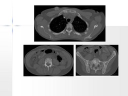
General Discussion:

For bone marrow metastases, overall sensitivity of PET and bone scan is comparable, but PET has superior specificity.FDG PET is superior to bone scan for detecting osteolytic metastases but is inferior for visualizing osteoblastic metastases. FDG PET cannot replace bone scan for detection of bone metastases in breast cancer. Patients with more aggressive tumors may benefit more from PET given the high likelihood of osteolytic bone metastases with those malignancies.

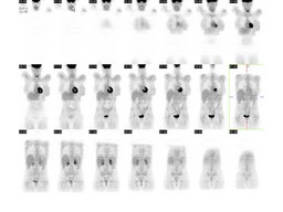
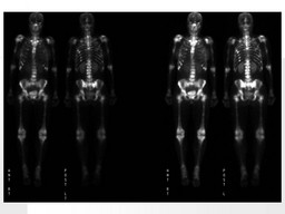
99 mTc-MDP shows the osteoblastic bone reaction to tumor infiltration, 18F-FDG measures glucose uptake into the tumor itself. This, along iwith is better resolution, allows PET to detect small volumes of disease before a secondary cortical bone reaction appears.  In a series of 25 patients with bone metastases (16 secondary to breast cancer and nine secondary to lung cancer) in whom both BS and PET procedures were performed before and after chemotherapy, BS and PET showed discordant results in five patients (20%) . These five patients showed improvement on PET examinations over time, with a reduction in FDG uptake and/or in the number of lesions detected. In three of them, BS suggested progression of metastatic disease and in the remaining two, results on serial BS remained unchanged. Clinical follow-up, serial tumor marker levels and radiological confirmation supported the PET findings. Therefore, in this preliminary experience, serial PET studies seemed more accurate than serial BS in treatment control of patients with bone metastases, in both types of primary tumor. It is important to know the danger of misinterpretation of BS due to the flare phenomenon and a persistent bone reaction lasting for more than 6–12 months after chemotherapy.

References: 1. Eugene C. Lin, Abass Alavi, et al. A clinical Guide PET and PET/CT; Page 112-116.
2. Takayoshi Uematsu, Sachiko Yuen, Seigo Yukisawa,et al. Comparison of FDG PET and SPECT for Detection of Bone Metastases in Breast Cancer. AJR 2005; 184:1266-1273
3. J. R. García,  M. Simó, M. Soler, et al. Relative roles of bone scintigraphy and positron emission tomography in assessing the treatment response of bone metastases. European Journal of Nuclear Medicine and Molecular Imaging© Springer-Verlag 2005
Comments:
No comments posted.
Additional Details:

Case Number: 106823Owner(s): Xiaoni Hong and Jerold Wallis, Assoc Prof of RadiologyLast Updated: 02-07-2013
Anatomy: Skeletal System   Pathology: Other
Modality: Nuc MedAccess Level: Readable by all users, writable by NucMed Certifiers
Keywords: widespread osseous metastises diseaseACR: 40000.12170

Case has been viewed 49 times.
Certified by Jerold Wallis on 06-17-2009

The reader is fully responsible for confirming the accuracy of this content.
Text and images may be copyrighted by the case author or institution.