After viewing the image(s), the Full history/Diagnosis is available by using the link here or at the bottom of this page
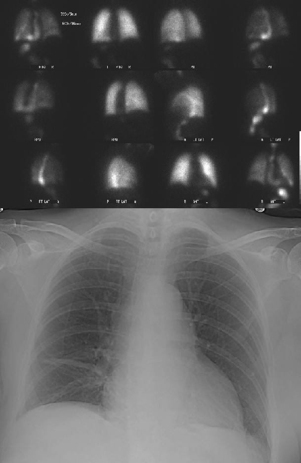
Upper study: First and fourth column are aerosol ventilation images. Second and third column are perfusion images. Ventilatory and perfusion images corresponding to the same projections are adjacent to each other. Lower study: Frontal chest radiograph performed the same day as the ventilation-perfusion examination.
View main image(vq) in a separate viewing box
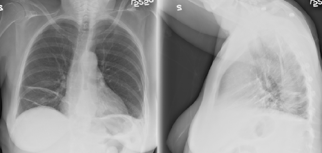
View second image(xr). PA and lateral chest radiographs performed two days prior to the ventilation-perfusion examination.
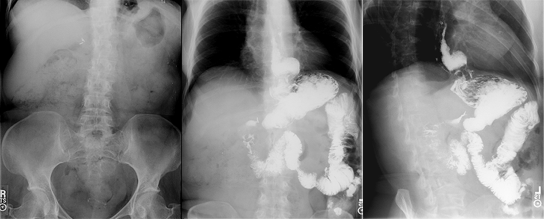
View third image(gs). Scout, frontal and left anterior oblique abdominal images from an upper gastrointestinal series performed one year prior to the ventilation-perfusion examination.
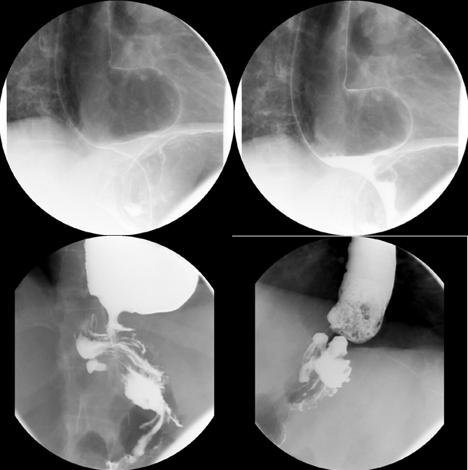
View fourth image(fl). Four select spot images of the gastroesophageal junction from same upper gastointestinal series examination.
Full history/Diagnosis is also available
Return to the Teaching File home page.