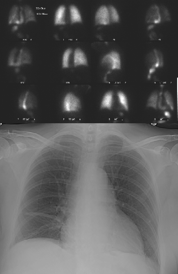

Upper study: First and fourth column are aerosol ventilation images. Second and third column are perfusion images. Ventilatory and perfusion images corresponding to the same projections are adjacent to each other. Lower study: Frontal chest radiograph performed the same day as the ventilation-perfusion examination.
View main image(vq) in a separate image viewer
View second image(xr). PA and lateral chest radiographs performed two days prior to the ventilation-perfusion examination.
View third image(gs). Scout, frontal and left anterior oblique abdominal images from an upper gastrointestinal series performed one year prior to the ventilation-perfusion examination.
View fourth image(fl). Four select spot images of the gastroesophageal junction from same upper gastointestinal series examination.
Full history/Diagnosis is available below
Her past surgical history is significant for repair of a hiatal hernia with subsequent Nissen fundoplication
Frontal chest radiograph: The heart size is at the upper limits of normal. There is atelectasis or scarring in both lung bases without significant change from a prior comparison study. No focal infiltrate, effusion or suspicious masses are identified.
PA and lateral comparison chest radiograph: The heart size is normal. There is atelectasis or scarring in the both lower lobes. Calcified left lung granulomas and an old healed left 7th rib fracture are noted. An air fluid level is noted in the middle mediastinum consistent with the known patulous esophagus.
Upper GI series: The scout radiograph demonstrates surgical clips consistent with cholecystectomy and a normal bowel gas pattern. Sutures in the lower mid pelvis are noted. The distal esophagus is dilated and there is pooling of contrast material and debris. There is delayed passage of the contrast agent through the gastroesophageal (GE) junction. The GE junction is below the diaphragm. A medially directed contrast-filled tongue like defect at the level of the GE junction is consistent with a fundoplication wrap. Additional images (not shown) demonstrated a normal stomach and duodenum with normal gastric emptying. No gastroesophageal reflux was elicited with provocative maneuvers. The small bowel follow-through examination was normal except for an incidentally noted proximal jejunal diverticulum.
The Tc-99m DTPA that reaches the alveoli passively diffuses across the alveolar epithelium and then the capillary endothelium to gain access to the circulatory system. The tracer is then cleared from the circulation via glomerular filtration. The pulmonary clearance of Tc-99m DTPA has a half-time of 60-90 minutes in normal patients. The rate of clearance is more rapid in smokers and in patients with disease that increase the permeability of the alveolar-capillary membrane, such as acute respiratory distress syndrome (ARDS).
Some Tc-99m DTPA aerosol is deposited in the oropharynx or is removed from the central airways via mucociliary transport, and then is swallowed and eliminated via the digestive tract. In this patient, swallowed Tc-99m DTPA has accumulated in the patuluous esophagus.
Parenthetically, nuclear medicine studies to evaluate esophageal obstruction, gastric emptying and gastroesophageal reflux may be performed using In-111 or Tc-99m labeled solid and liquid food. When a dual-tracer method is performed (In-111 DTPA in liquid food is used in conjunction with Tc-99m sulfur colloid-labeled solid food), liquid and solid emptying can be evaluated independently.
REFERENCES: Goodin, LJ (2000, March 19) Radiopharmaceutical Fact Sheet: Technetium (Tc-99m) Pentetate. Retrieved January 28, 2004, from Nuclear Medicine Technology Resource WebSite: http://www3.sympatico.ca/lgoodin/rp-4-0.htm
Williams, SC (2001) Nuclear Medicine Online Reference Text. Ventilation/Perfusion Imaging: Technique & Radiopharmaceuticals. Retrieved January 28, 2004, from Aunt Minnie Web Site: http://www.auntminnie.com/default.asp?sec=ref&sub=ncm&pag=pul&itemid=54282
Williams, SC (2001) Nuclear Medicine Online Reference Text. Inflammatory Lung Disease: Aerosolized Tc-99m DTPA Evaluation. Retrieved January 28, 2004, from Aunt Minnie Web Site: http://www.auntminnie.com/default.asp?sec=ref&sub=ncm&pag=pul&itemid=55011&d=1
Graber, J et al (2002, January) Laparoscopic Gastric Fundoplication for Treatment of Gastroesophgaeal Reflux Disease. Minnesota Medicine, Vol 85. Retrieved February 9, 2004, from Minnesota Medicine publications: http://www.mmaonline.net/publications/MNMed2002/January/Graber.html
References and General Discussion of Ventilation Perfusion Scintigraphy (Anatomic field:Lung, Mediastinum, and Pleura, Category:Organ specific)
Return to the Teaching File home page.