After viewing the image(s), the Full history/Diagnosis is available by using the link here or at the bottom of this page

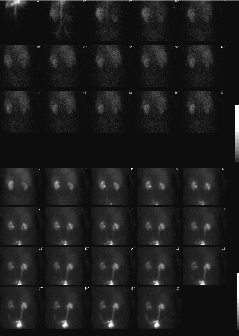
Top images: Posterior projection images from renal angiogram. Bottom images: Additional sequential posterior projection images through 20 minutes following administration of radiopharmaceutical.
View main image(rs) in a separate viewing box
View second image(rs). Top images: Posterior projection images for 22 minutes following administration of Lasix. Bottom right image: posterior static image, post Lasix, post void.
View third image(rs). Top images: Split renal function analysis curve. Bottom image: Post Lasix renal function analysis curve
Full history/Diagnosis is also available
Return to the Teaching File home page.