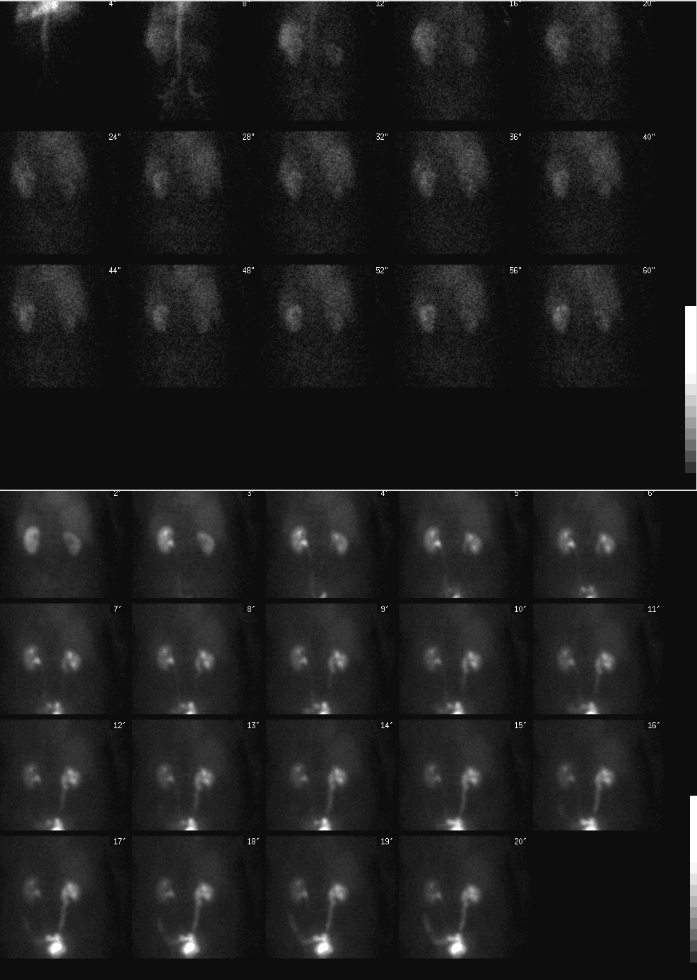

Top images: Posterior projection images from renal angiogram. Bottom images: Additional sequential posterior projection images through 20 minutes following administration of radiopharmaceutical.
View main image(rs) in a separate image viewer
View second image(rs). Top images: Posterior projection images for 22 minutes following administration of Lasix. Bottom right image: posterior static image, post Lasix, post void.
View third image(rs). Top images: Split renal function analysis curve. Bottom image: Post Lasix renal function analysis curve
Full history/Diagnosis is available below
Sequential renal images through 20 minutes: The left kidney is of normal size with normal uptake and excretion of tracer. The right kidney is small in size but maintains its reniform shape. There is mildly delayed excretion of tracer from the right kidney. Relatively decreased activity centrally in the right kidney corresponds to a dilated pelvicalyceal system. There is, however, progressive accumulation of tracer in the right collecting system. The right ureter is mildly enlarged. The left ureter is not completely visualized. The bladder is unremarkable.
Split renal curve analysis: The left renal function curve is normal. The right renal function curve indicates decreased renal function. The right kidney contributes 34% and the left kidney 66% of total renal function.
Sequential renal images post lasix: To evaluate for obstruction, 40 mg of furosemide was administered via slow intravenous injection approximately 30 minutes after the start of the examination. Sequential images were obtained for an additional 22 minutes. There is prompt clearance of residual pelvicalyceal activity from the left collecting system after diuretic administration. There is slow clearance of activity from a dilated right pelvicalyceal system and some clearance of activity from the right ureter.
Progressive tracer accumulation is noted in the lower pelvis extending to the left hemicolon (lateral to to left ureter). This is consistent with the known colovesicular fistula.
Lasix renal curve analysis: There is normal clearance of activity in the left kidney following lasix administration. The half time tracer clearance from the left collecting system is normal, 4 minutes. There is slow clearance of activity from the right collecting system and ureter. There is a prolonged half time tracer clearance from the right collecting system, 41 minutes. This supports the imaging findings of partial obstruction.
Lasix is a loop diuretic that inhibits reabsorption of electrolytes in the ascending limb of the loop of Henle, proximal and distal tubules. This results in an increase in excretion of water, sodium and chloride. Rapid emptying of the collecting system with a steep declining curve indicates dilation without obstruction. Whereas, slow emptying indicates partial obstruction or renal dysfunction with inability to respond to diuresis.
Adequate hydration prior to the start of the renal scintigraphy examination is important when evaluating renal function.
See case RS029 for a companion case of renal obstruction.
Colorectal fistulas can be caused by several different disease processes. The most common cause in the U.S. is from diverticulitis. Other common causes include inflammatory bowel disease (e.g. Crohn's disease or ulcerative colitis) and malignancy (colon or cervical). Colovesicular fistulas may also form from following radiation therapy. In this patient, the colovesicular fistula is presumed secondary to her prior radiation therapy although she also has colonic diverticulosis.
REFERENCES: Krco MJ et al. "Colovesical fistulas." Urology. 1984. Apr: 23(4):340-2. Griffeth, LK. "Pharmacologic Interventions in Nuclear Medicine." 2004. Mallinckrodt Institute of Radiology. March 8, 2004.
The single LPO view of the abdomen following administration of the hypaque contrast demonstrates filling of the urinary bladder. Note the outline of the rectal tube balloon as well as the outline of the foley catheter balloon by contrast indicating the presence of a colovesicular fistula. The fistula tract could not be clearly delinated due to overlap from the hip arthroplasty. There are changes consistent with diverticulosis in the distal sigmoid colon.
A percutaneous right nephrostomy tube was initially placed. An antegrade nephrostogram subsquently was performed (images not included)demonstrating a dilated right renal collecting system without filling defects. A few centimeters distal to the right ureteropelvic junction, a short segement of high grade stenosis with associated sharp angulation was noted. A long segment of stenosis in the distal ureter was noted approximately 6-8 cm in length, ending at the ureteropelvic junction.
The patient was brought to the operating room where she underwent repair of the colovesicular fistula, resection of a small bowel stricture of the terminal ileum/cecum and distal diseased sigmoid colon and rectum, and formation of a colostomy and closure of the rectal stump. A left ureteral stent was also placed. Pathology revealled a fistulous tract in the rectosigmoid colon with changes consistent with serositis. Pathology evaluation of the terminal ileum and cecum revealled well differentiated invasive adenocarcinoma. Surgical margins were free of tumor and zero of fifteen lymph nodes removed were positive for malignancy. Angioplasty and stenting of the distal right ureteral stricture is anticipated following the patient's recovery from her colovesicular fistula repair.
View followup image(gs). Scout and LPO views of the abdomen from a Hypaque contrast enema examination
References and General Discussion of Renal Scintigraphy (Anatomic field:Genitourinary System, Category:Organ specific)
Return to the Teaching File home page.