After viewing the image(s), the Full history/Diagnosis is available by using the link here or at the bottom of this page
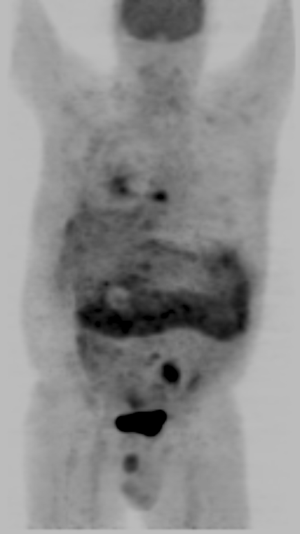
Whole-body FDG-PET images demonstrate increased activity along a prominent, thickened omentum as well as other foci of uptake later shown to represent skeletal metastases in the left chest wall, in the left iliac wing, and in the T8, T10, and L5 vertebrae.
View main image(pt) in a separate viewing box
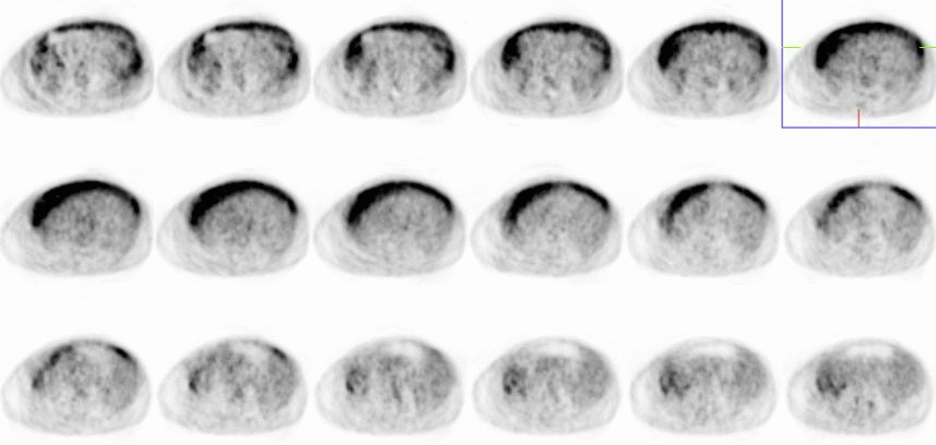
View second image(pt). Axial images confirm thickening and dramatically increased uptake in the greater omentum, consistent with omental caking.
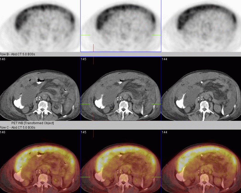
View third image(pt). The fused PET/CT images demonstrate that the omental caking seen on CT scan demonstrates markedly increased PET activity.
Full history/Diagnosis is also available
Return to the Teaching File home page.