Case Author(s): Jabi Shriki, M.D. and Barry A. Siegel, M.D. , 9/12/05 . Rating: #D2, #Q3
Diagnosis: Omental caking due to metastatic non-small cell lung carcinoma
Brief history:
62 year-old man with prior right pneumonectomy for non-small cell lung cancer. A prior CT scan detected nodularity and enhancement along the right side of the omentum.
Images:

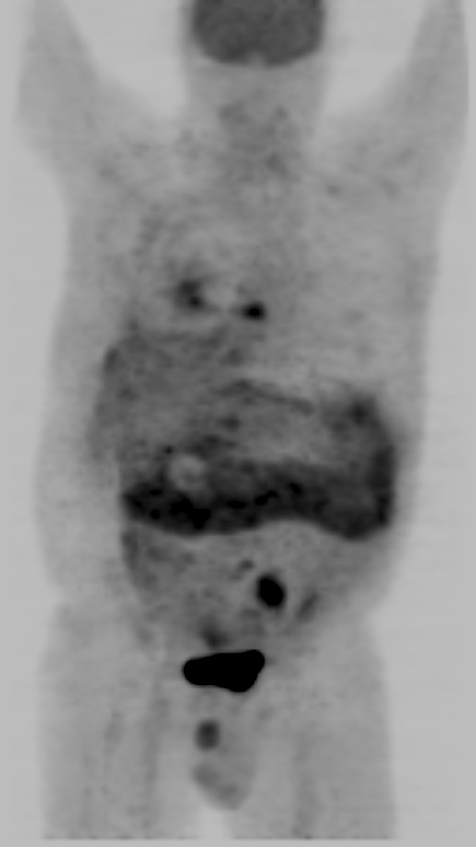
Whole-body FDG-PET images demonstrate increased activity along a prominent, thickened omentum as well as other foci of uptake later shown to represent skeletal metastases in the left chest wall, in the left iliac wing, and in the T8, T10, and L5 vertebrae.
View main image(pt) in a separate image viewer
View second image(pt).
Axial images confirm thickening and dramatically increased uptake in the greater omentum, consistent with omental caking.
View third image(pt).
The fused PET/CT images demonstrate that the omental caking seen on CT scan demonstrates markedly increased PET activity.
Full history/Diagnosis is available below
Diagnosis: Omental caking due to metastatic non-small cell lung carcinoma
Full history:
The patient is a 62 year-old man with a primary diagnosis of adenocarcinoma of the right lung, which was resected 11 months prior to the PET scan. The patient underwent a CT scan with contrast (not shown) prior to the PET scan, which demonstrated that there was soft tissue thickening, increased attenuation, and nodularity along the greater omentum. Unfortunately, he was lost to follow-up at that time. Subsequently, he re-presented for evaluation of abdominal pain. The PET/CT obtained for restaging at that time demonstrated gross thickening of the omentum. Ultrasonography-guided biopsy (not shown) obtained tissue felt to be consistent with metastatic adenocarcinoma.
Radiopharmaceutical:
14.9 mCi F-18 fluorodeoxyglucose
Findings:
There is dramatically increased uptake along the greater omentum. This correlates with thickening and nodularity seen on the CT scan consistent with omental caking. Additionally, skeletal metastases are seen in the left chest wall, in the left iliac wing, and in the T8, T10, and L5 vertebrae.
Discussion:
Omental caking is a term reserved for metastatic disease along the greater omentum, which usually ensues as a result of peritoneal spread of tumor. The common malignancies that spread via peritoneal seeding, and thus cause omental caking include ovarian, gastric, colonic, and pancreatic cancers. Other more rare causes of omental caking include renal cell carcinoma and primary mesothelioma of the peritoneum. To our knowledge, metastatic lung carcinoma with resultant metastatic omental caking has not been reported.
Differential Diagnosis List
Differential Diagnosis for Omental Caking:
1. GI carcinoma from a colonic or gastric primary.
2. Ovarian carcinoma.
3. Pancreatic carcinoma.
4. Rarely renal carcinoma.
5. Primary peritoneal mesothelioma.
6. Tuberculous peritonitis.
ACR Codes and Keywords:
References and General Discussion of PET Tumor Imaging Studies (Anatomic field:Gasterointestinal System, Category:Neoplasm, Neoplastic-like condition)
Search for similar cases.
Edit this case
Add comments about this case
Return to the Teaching File home page.
Case number: pt141
Copyright by Wash U MO

