
After viewing the image(s), the Full history/Diagnosis is available by using the link here or at the bottom of this page
Coronal (above) and axial (below) F18-FDG PET images
View main image(pt) in a separate viewing box
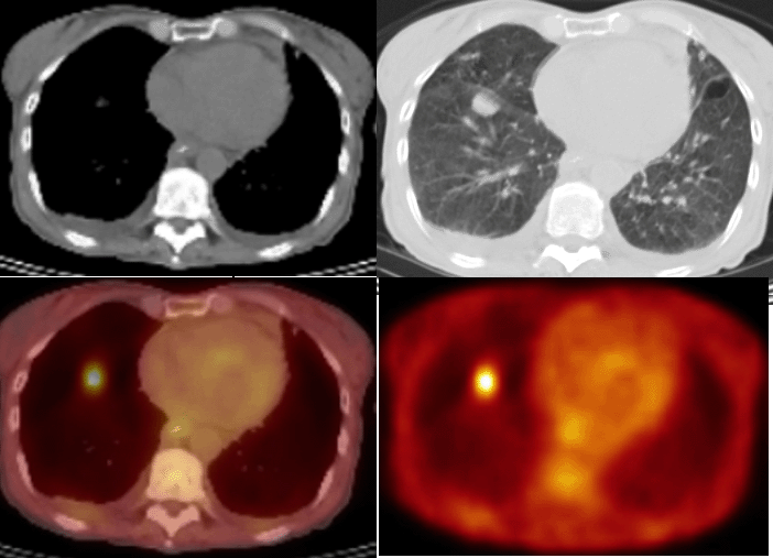
View second image(pt). Axial CT in soft tissue (upper left) and lung windows (upper right); fused PET-CT (lower left) and F18-FDG PET (lower right) images.
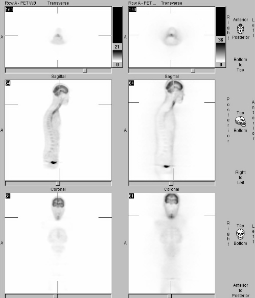
View third image(pt). Attenuated-corrected and non-attenuation corrected PET images.
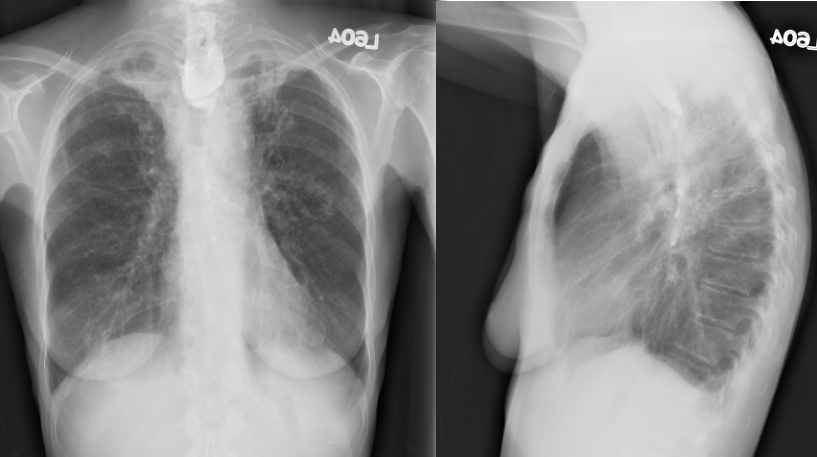
View fourth image(xr). PA and lateral chest radiographs performed the same day as the PET examination.
Full history/Diagnosis is also available
Return to the Teaching File home page.