Case Author(s): Dennis Hsueh, M.D., Dallas Sorrell, M.D. and Farrokh Dehdashti, M.D. , 08/13/03 . Rating: #D3, #Q3
Diagnosis: Attenuation-Correction Artifact on FDG-PET/CT due to retained barium in a esophago-retropharyngeal fistulous tract
Brief history:
Sixty-year-old woman with a history of supraglottic laryngeal carcinoma treated surgically and with radiation therapy. Follow-up CT images of the neck and chest demonstrated a new pulmonary nodule. PET requested to characterize this nodule.
Images:

Coronal (above) and axial (below) F18-FDG PET images
View main image(pt) in a separate image viewer
View second image(pt).
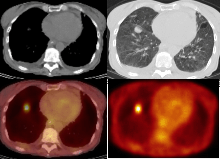
Axial CT in soft tissue (upper left) and lung windows (upper right); fused PET-CT (lower left) and F18-FDG PET (lower right) images.
View third image(pt).
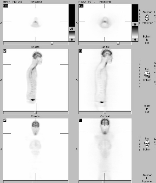
Attenuated-corrected and non-attenuation corrected PET images.
View fourth image(xr).
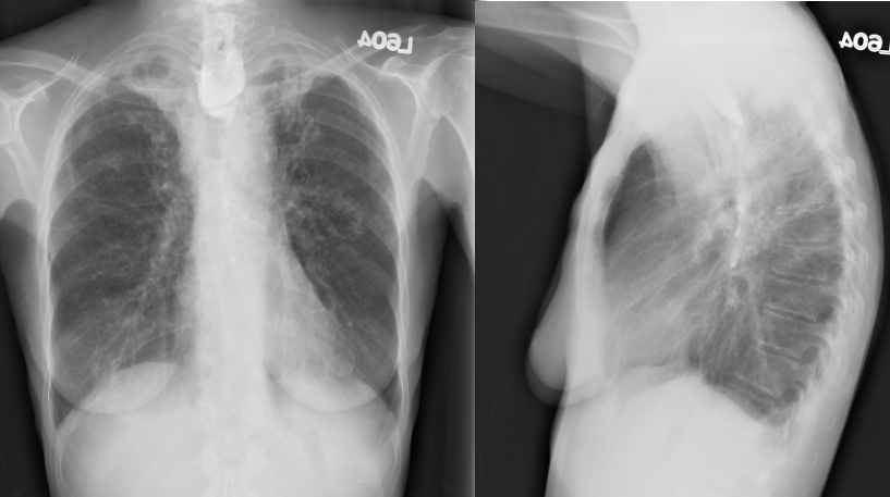
PA and lateral chest radiographs performed the same day as the PET examination.
Full history/Diagnosis is available below
Diagnosis: Attenuation-Correction Artifact on FDG-PET/CT due to retained barium in a esophago-retropharyngeal fistulous tract
Full history:
This is a 60-year-old woman with a history of supraglottic laryngeal carcinoma treated with laryngectomy and radiation therapy three years prior to her current examination. Her post therapy course has been complicated by esophageal strictures requiring esophagoscopy and dilatation and by recurrent fistulas. A new pulmonary nodule was identified on CT chest examination obtained during recent evaluation of esophagus. An FDG-PET study was requested to evaluate for metabolic activity in this nodule.
Radiopharmaceutical:
F-18 fluorodeoxyglucose (FDG)
Findings:
PET images: Retained barium within a false esophageal lumen results in artifactual increased activity, only appreciated on the attenuation-corrected images. Intense increased FDG activity is associated with the right lower lobe pulmonary nodule. Measured maximum standardized uptake value is 6.1 which is suspicious for malignancy. Mild diffuse FDG uptake is noted at the laryngectomy site and bilateral upper lobes consistent with post-operative and post-radiation changes. Minimal FDG uptake associated with posterior pleural thickening, likely due to scarring.
Chest radiograph (not shown, obtained same day as PET examination): Demonstrates retained barium in the proximal third of the esophagus or false lumen.
Discussion:
Contemporary PET examinations utilize reconstruction of algorithms for attenuation correction. That is, a correction factor is applied to the emission data to account for differences in thickness and path length of emitted photon. Photons from shallow (e.g. skin) or gas containing structures (e.g. lungs) are attenuated less; whereas, those in deep soft tissue structures are attenuated more. Advantages with attenuation correction include less image distortion and easier anatomic localization.
Conventional PET scanners generate an attenuation map by obtaining transmission images with the patient positioned between a rotating germanium source and the camera detectors. With current dual modality PET/CT scanners, the CT data substitutes for the PET transmission data. CT acquisition not only is more rapid but also generates less image noise compared with the PET transmission scan. Thus, total scan duration is reduced, and with image fusion, anatomic localization is markedly improved.
Because of differences in photon energy during the acquisition of CT data (40-140 keV) compared with PET data (511 keV), the CT attenuation data must be transformed into linear attenuation coefficients at 511 keV. Current attenuation maps have a maximum threshold of approximately 300 Hounsfield units. Difficulties arise when very dense objects (e.g. metal, barium, dental amalgam) are encountered because attenuation of 40-140 keV photons is greater than with 511 keV photons. This results in an over-estimation of attenuation and corrected images appear to demonstrate increased activity.
Followup:
The patient underwent a CT guided biopsy of the right lower lobe mass. Due to difficulties during the biopsy (the patient developed hemoptysis), the samples collected were non-diagnostic.
A follow up CT chest examination 3 months later demonstrated interval enlargement of the pulmonary nodule from 1.8 x 1.4 cm to 2.3 x 1.9 cm likely representing pulmonary metastatic disease.
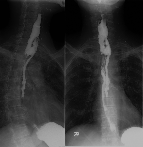
View followup image(gs).
Barium swallow examination performed 2 days prior to PET imaging examination. This demonstrates a fistula between the esophagus and the retropharyngeal soft tissues.
Major teaching point(s):
1. When reviewing PET/CT with CT based corrected attenuation images, examination of the non-attenuation corrected images and scout image is essential to avoid misleading artifacts.
2. One potential solution to this problem is to raise the maximum threshold when deriving the attenuation map so that when differences in attenuation of very dense objects are encountered, it is properly included in the correction.
Differential Diagnosis List
If the findings had persisted on non-attenuated corrected images, esophagitis or less likely, diffuse mucosal or mural neoplasm of the esophagus would also be considered.
REFERENCES:
Bujenovic, S. et al. "Artifactual 2-Deoxy-2(F18)Fluoro-D-Glucose Localization Surrounding Metallic Objects in a PET/CT Scanner Using CT-Based Attenuation Correction." Molecular Imaging and Biology. Vol. 5. No. 1. 20-22, 2003.
Seigel, B. “Interpreting Oncologic FDG PET Images.” PET Imaging for the Radiologist. CD-ROM. ACR Publications. September 2002.
ACR Codes and Keywords:
- General ACR code: 69
- Lung, Mediastinum, and Pleura:
6.12163 "PET"
References and General Discussion of PET Tumor Imaging Studies (Anatomic field:Lung, Mediastinum, and Pleura, Category:Other(Artifact))
Search for similar cases.
Edit this case
Add comments about this case
Return to the Teaching File home page.
Case number: pt102
Copyright by Wash U MO