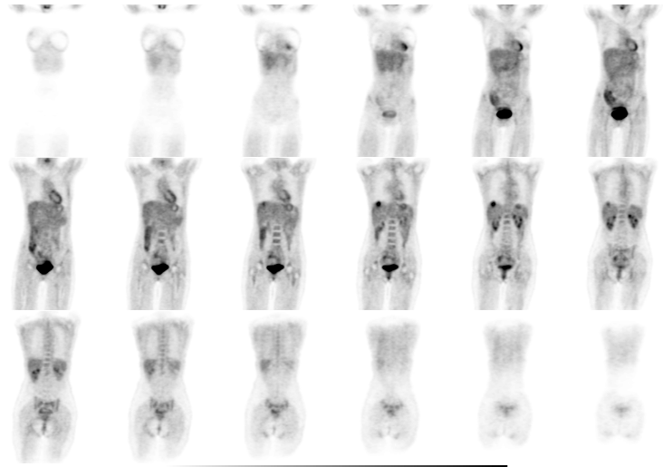
Coronal whole body images
View main image(pt) in a separate image viewer
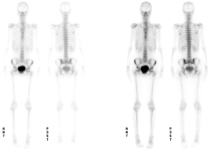
View second image(bs). Dual intensity anterior and posterior whole body images of a bone scan.
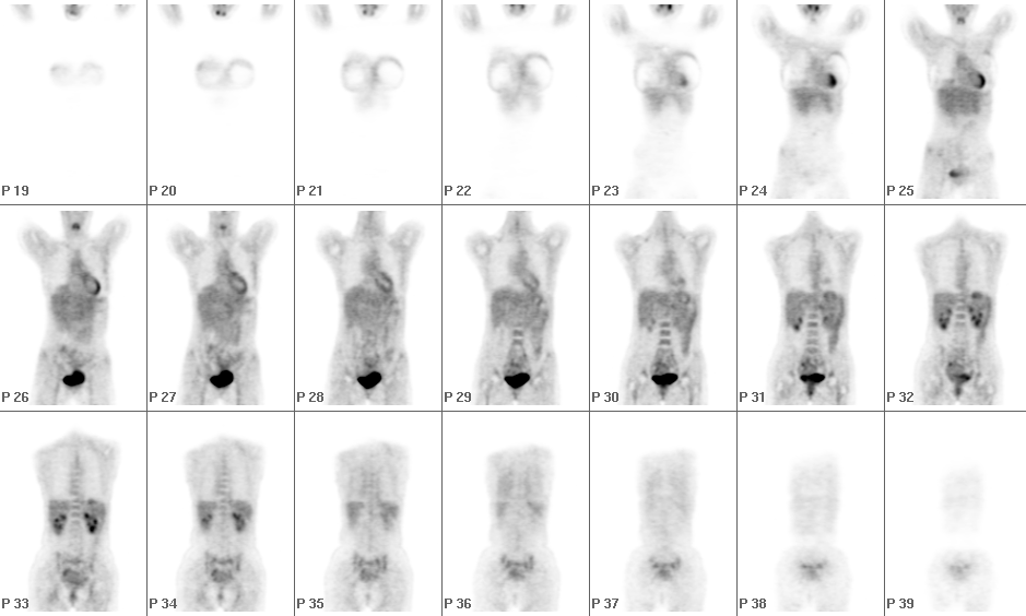
View third image(pt). Coronal whole body images from a study one year previously
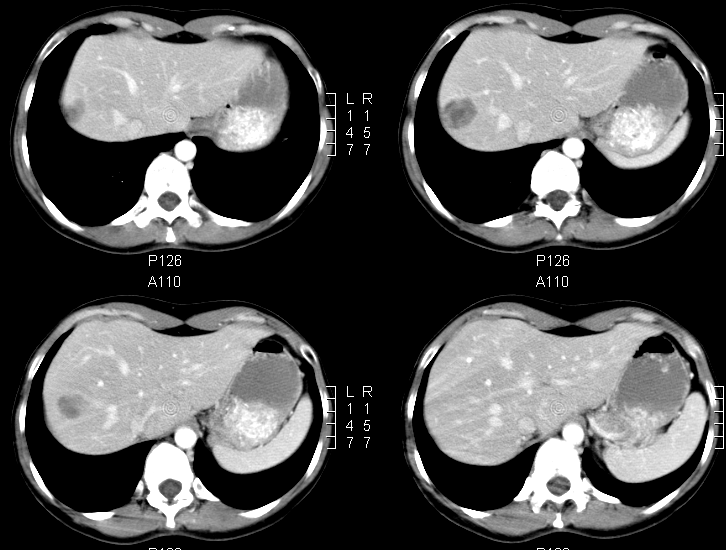
View fourth image(ct). Selected axial images from a recent contrast-enhanced CT examination
Full history/Diagnosis is available below
Current Bone Scan: Mild increased uptake in the left 4th and 6th ribs. These were decreased compared with a prior bone scan and were felt most consistent with healing rib fractures.
Prior PET Study (one year prior): Bilateral breast implants were noted, but there was no abnormal uptake to suggest residual or recurrent tumor.
Current CT Study: Low attenuation lesion in the posterior segment of the right hepatic lobe, highly suspicious for a metastasis.
Recurrence of breast cancer is often local at the excision site. Lymphatic spread to the axilla is the most common route of regioal metastatic spread. Distant metastatic spread is most common to the bones, liver, lungs and brain.
PET is useful in staging and restaging in breast carcinoma as well as for assessing response to therapy. PET may also detect bone metastases that are not seen on bone scan as evidenced in another teaching file on this site (MIR Teaching file case pt057).
Sclerotic metastases in breast cancer may not bevisualized by PET, while lytic breast metastases are more frequently seen on PET.
References and General Discussion of PET Tumor Imaging Studies (Anatomic field:Breast, Category:Neoplasm, Neoplastic-like condition)
Return to the Teaching File home page.