Case Author(s): Yungao Ding, MD, PhD and Henry Royal, MD , 5/11/2001 . Rating: #D., #Q.
Diagnosis: Metastatic Breast Cancer
Brief history:
50 year old female with newly diagnosed left breast cancer
Images:

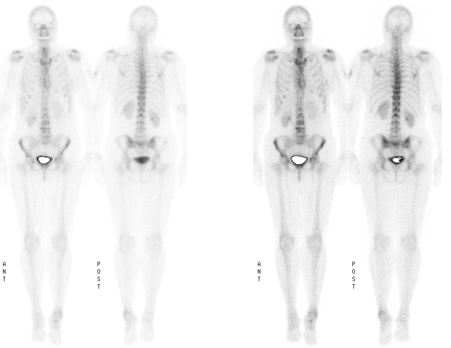
Whole-body bone scintigraphy
View main image(bs) in a separate image viewer
View second image(pt).
Posterior 3-D rendered PET image
View third image(pt).
Saggital PET image through the spine
Full history/Diagnosis is available below
Diagnosis: Metastatic Breast Cancer
Full history:
50 year old female with newly diagnosed left breast cancer.
Radiopharmaceutical:
19.0 mCi Tc-99m MDP, i.v.
Findings:
There was no definite evidence of bone metastasis on bone scan. The radiotracer uptake in the soft tissue of left upper arm was due to injection granuloma for allergy medication. The increased uptake at the superior end plate of L4 was compatible with degenerative change. The minimally increased uptake in the L2 spinous process was thought to be positioning. Post-traumatic change was noted in left ankle according to patient's history. The tracer uptake in the region of ethmoid sinus is consistent with sinusitis.
The patient subsequently underwent left mastectomy with left axillary node dissection. There were metastases involving multiple left axillary lymph node. Therefore, systemic metastasis was strongly suspected clinically. CT of the chest, abdomen and pelvis was performed following mastectomy and showed no evidence of metastasis both in skeletal and non-skeletal systems. Tumor PET imaging was then performed.
PET with 15 mCi F-18 FDG showed abnormal uptake of FDG involving lower cervical, thoracic and lumbar spine, pelvis including sacrum, ischia and right iliac bone, consistent with widespread bone metastasis. There was also mild uptake of FDG in the proximal left femur probably due to metastasis. The mild uptake in the right shoulder and sternoclavicular joints were consistent with degenerative changes. Post-operative changes were noted in the left chest and axilla.
Discussion:
Bone scan is routinely performed for patients with breast cancer, and it is generally regarded as a sensitive study for bone metastasis. However, there is no "gold standard" to really verify the sensitivity of bone scan for systemic bone metastasis from breast cancer. One can not biopsy every bone/bone marrow, or every part of a single bone. Bone scan detects new bone formation/bone turnover at the site of metastases. In the early stage of metastasis when the metastasis is still mainly confined to marrow without significant new bone formation/bone turnover, conceivably bone sacn will not be sensitive to detect metastsis as demonstrated in this case. However, PET detects metastasis based on increased glucose metabolism regardless of new bone formation/bone turnove. Unlike bone scan, PET also detect soft tissue metastasis. Therefore, for patients with strong clinical suspicion of metastasis PET is probably the study of choice to evaluate both skeletal and non-skeletal metastasis.
Major teaching point(s):
Depending on the amount of new bone formation/bone turnover, bone scan may or may not be sensitive enough to detect early bone metastasis when metastasis is already obvious on PET.
ACR Codes and Keywords:
References and General Discussion of PET Tumor Imaging Studies (Anatomic field:Skeletal System, Category:Neoplasm, Neoplastic-like condition)
Search for similar cases.
Edit this case
Add comments about this case
Return to the Teaching File home page.
Case number: pt057
Copyright by Wash U MO

