After viewing the image(s), the Full history/Diagnosis is available by using the link here or at the bottom of this page

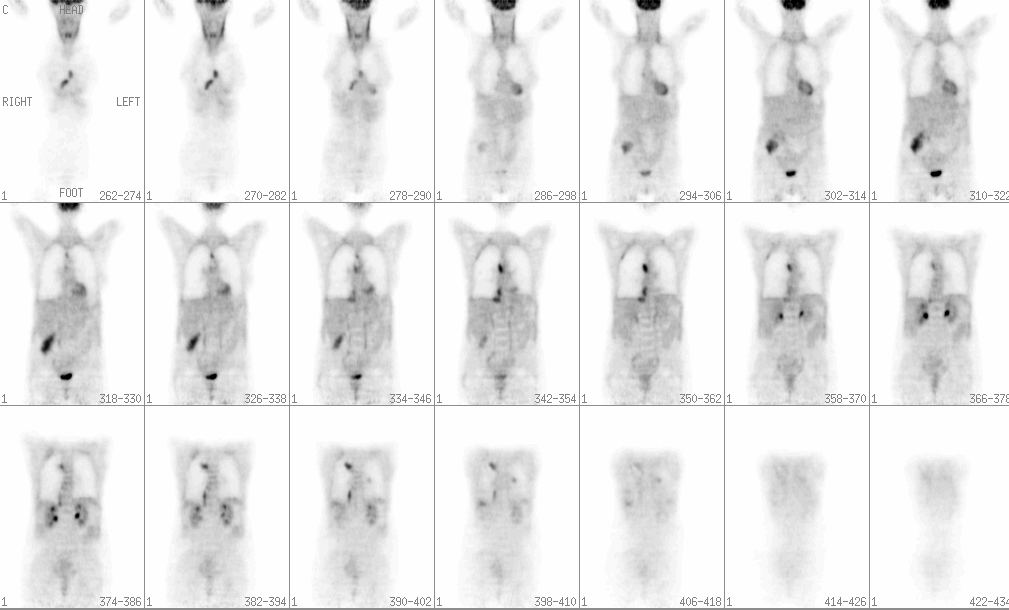
Coronal PET images from the skull base to proximal thighs.
View main image(pt) in a separate viewing box
View second image(pt). Anterior, RAO, LAO, posterior reprojection images, and coronal, sagittal and axial planar images at various levels.
View third image(ct). Axial CT images of the upper and mid thorax.
View fourth image(ct). Axial CT images of the lower thorax and abdomen.
Full history/Diagnosis is also available
Return to the Teaching File home page.