Case Author(s): Gabriel De Simon, M.D. and Barry A. Siegel, M.D. , 02/01/01 . Rating: #D3, #Q4
Diagnosis: Talc pleurodesis simulating pleural metastases on FDG PET imaging.
Brief history:
Severe emphysema with several episodes of spontaneous pneumothorax, treated by pleurodesis. The patient also has a cecal mass suspicious for malignancy, on an abdominal CT scan.
Images:
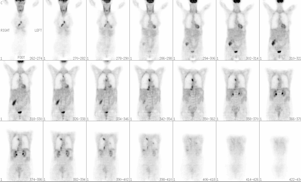
Coronal PET images from the skull base to proximal thighs.
View main image(pt) in a separate image viewer
View second image(pt).
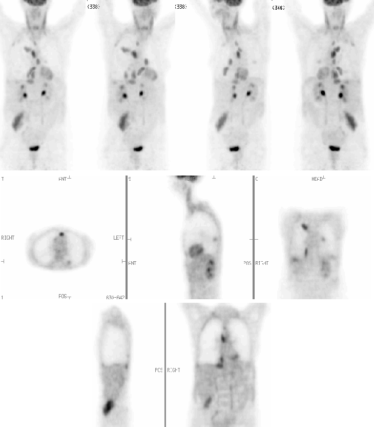
Anterior, RAO, LAO, posterior reprojection images, and coronal, sagittal and axial planar images at various levels.
View third image(ct).
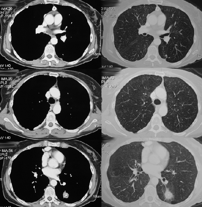
Axial CT images of the upper and mid thorax.
View fourth image(ct).
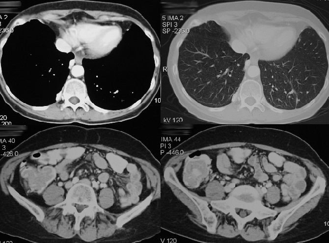
Axial CT images of the lower thorax and abdomen.
Full history/Diagnosis is available below
Diagnosis: Talc pleurodesis simulating pleural metastases on FDG PET imaging.
Full history:
66-year-old woman with known severe emphysema due to smoking, who had several spontaneous right pneumothoraces, treated by chest tube placement and talc pleurodesis, approximately one year prior to the PET study. A CT scan of the chest, abdomen and pelvis performed prior to the PET scan demonstrated a lower lobe pulmonary mass suspicious for a primary bronchogenic cancer, and a mass in the cecum suggestive of a colonic neoplasm.
Radiopharmaceutical:
3.2 mCi F-18 Fluorodeoxyglucose i.v.
Findings:
WHOLE BODY TUMOR PET IMAGING:
1. Curvilinear areas of intensely increased FDG uptake on the pleural reflection of the right hemithorax, located predominantly on the mediastinal surface. Most of the increased uptake is on the inferior paravertebral pleural surface just above the right hemidiaphragm, and extends cephalad to involve more apical regions of the paravertebral pleura.
2. Focal region of moderately increased activity, with standardized uptake value of 1.8, in the posterior left lower lobe. FDG uptake is eqivocal for malignancy.
3. Midline anterior mediastinal focal uptake, just above the base of the ascending aorta.
3. Lobulated, intensely increased uptake in the cecum and proximal ascending colon, consistent with a colonic neoplasm. No features of abdominal lymphadenopathy or hepatic metastases.
CONTRAST-ENHANCED CT SCAN OF THE CHEST AND ABDOMEN:
1. Multiple right pleural-based masses with regions of focal high attenuation suggestive of calcium.
2. Left lower lobe spiculated pulmonary mass with central calcification, measuring approximately 2.5 cm, consistent with a primary bronchogenic carcinoma.
3. Circular calcification in the anterior mediastinum, just above the base of the ascending aorta.
3. 5-cm, enhancing mass in the cecum and ascending colon, likely a colonic malignancy.
Discussion:
Talc, a soft granular mineral composed of an acid metasilicate of magnesium (H2 Mg3 {SiO3}4), is more effective for pleurodesis than tetracyclines, quinacrine, mustine or bleomycin, with a reported success rate of approximately 91%. Talc was administered in this patient with emphysema subsequent to several recurrent spontaneous pneumothoraces, as a slurry by chest tube, twelve months prior to the FDG PET scan. Pathological studies have demonstrated that talc incites an intense granulomatous pleural inflammatory reaction, usually occurring within 24 hours, and often persisting many months. Although the patient has a suspicious primary cecal malignancy by CT, substantiated by intensely increased FDG uptake, pleural inflammation due talc pleurodesis is presumed to be the cause of increased pleural activity rather than metastatic deposits, because the FDG distribution correlates almost exactly with that of the high density talc deposits on the CT scan. In addition, it would be very unlikely for colonic cancer metastases to involve the pleura so extensively on the right side without contralateral involvement, and metastases with calcification would likely have arisen from a mucinous colonic primary neoplasm, with tumoral low cellular density and mucin production expectedly resulting in less intense FDG activity than present in the cecal lesion or pleural surface of the patient. The increased use of FDG PET to diagnose and stage thoracic malignancy, the usefulness of whole body imaging for staging neoplasms, and the increasing use of talc for pleurodesis, may result in more cases in which regions of intense pleural uptake caused by the inflammatory response to talc, are misinterpreted as malignancy. Correlation with the CT chest is mandatory to avoid diagnostic errors in patients with a known history of talc pleurodesis. The patient also has a left lower lobe mass with standardized uptake value of 1.8, which although equivocal for malignancy by FDG uptake, remains highly suspicious for a second primary bronchogenic cancer given the spiculated appearance on CT and backround of severe emphysematous changes.
Reference: Murray JG, Erasmus JJ, Bahtiarian EA and Goodman PC. Talc pleurodesis simulating pleural metastases on F18 FDG PET imaging. AJR 1997; 168:359-360.
Followup:
The patient underwent resection of a 23 cm segment of large intestine, with histological examination demonstrating multiple tubular villous adenomas, the largest measuring 9.8 cm, with focal high grade dysplasia but no evidence of malignancy or invasion at the resection margins. PET is not sufficiently specific in the differentiation of highly dysplastic villous adenomas and carcinomas, and the intense FDG uptake in the cecum is not surprising for this benign tumor. The patient was to undergo resection of the left lower lobe mass, but refused surgery and will thus have followup CT scans four monthly for re evaluation. A repeat CT scan performed on 2/23/01 demonstrated no interval growth or change in morphology of the left lower lobe mass.
ACR Codes and Keywords:
References and General Discussion of PET Tumor Imaging Studies (Anatomic field:Lung, Mediastinum, and Pleura, Category:Inflammation,Infection)
Search for similar cases.
Edit this case
Add comments about this case
Read comments about this case
Return to the Teaching File home page.
Case number: pt050
Copyright by Wash U MO