After viewing the image(s), the Full history/Diagnosis is available by using the link here or at the bottom of this page
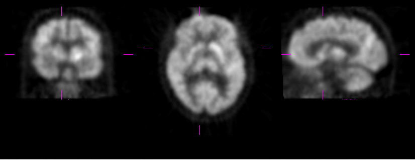
Selected coronal, axial and sagittal images of brain FDG-PET study.
View main image(pt) in a separate viewing box
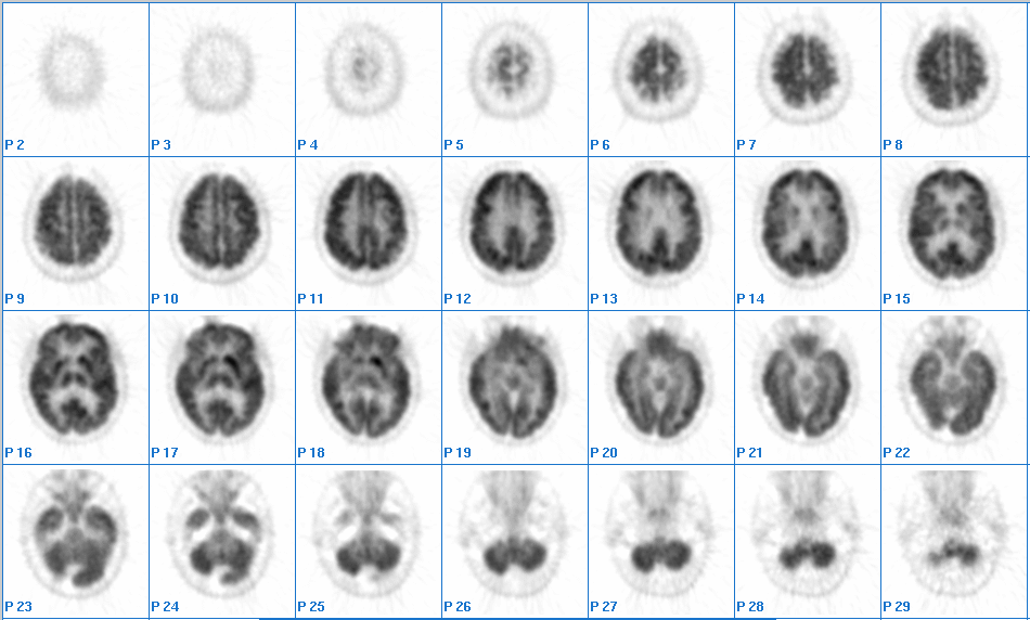
View second image(pb). Sequential axial images from brain FDG-PET study
View third image(ct). Axial CT image at the level of basal ganglia.
Full history/Diagnosis is also available
Return to the Teaching File home page.