Case Author(s): Bart Rydzewski, MD,Ph.D., Jerold Wallis, MD , 12/16/00 . Rating: #D3, #Q3
Diagnosis: Lacunar infarction of the right caudate nucleus.
Brief history:
73 y/o male being evaluated for a pulmonary nodule.
Images:
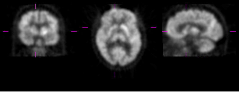
Selected coronal, axial and sagittal images of brain FDG-PET study.
View main image(pt) in a separate image viewer
View second image(pb).
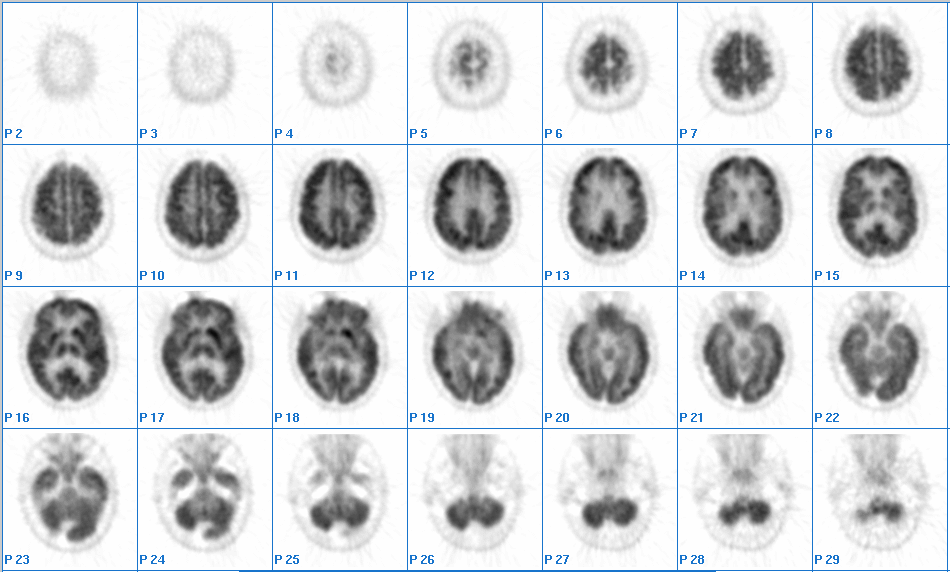
Sequential axial images from brain FDG-PET study
View third image(ct).
Axial CT image at the level of basal ganglia.
Full history/Diagnosis is available below
Diagnosis: Lacunar infarction of the right caudate nucleus.
Full history:
73 year old male smoker who presents for characterization of a right apical 1.7 x 2.3 cm ill-defined lung nodule and multiple subcentimeter mediastinal lymph nodes. Brain imaging study is performed as a part of a research protocol.
Radiopharmaceutical:
15.0 mCi F-18 Fluorodeoxyglucose i.v.
Findings:
Additional images of the brain were obtained (using the same tracer dose), under a research protocol, and without additional charge to the patient. These consisted of a standard (47-slice) emission PET imaging of the brain with a mathematical attenuation correction. The PET images demonstrate moderately asymmetric activity within the basal ganglia, right less than left, corresponding to a right caudate infarct on CT. No additional hypo or hypermetabolic lesions are identified to suggest metastatic disease.
Discussion:
Although, FDG-PET is not considered a primary imaging modality for cerebrovascular accidents, incidental cerebral infarctions will present on FDG-PET studies as focal regions of decreased metabolism. Brain FDG-PET studies must be compared with a recent cross-sectional brain imaging studies, to increase accuracy of interpretation of the FDG-PET findings.
ACR Codes and Keywords:
References and General Discussion of PET Tumor Imaging Studies (Anatomic field:Skull and Contents, Category:Metabolic, endocrine, toxic)
Search for similar cases.
Edit this case
Add comments about this case
Return to the Teaching File home page.
Case number: pt044
Copyright by Wash U MO