After viewing the image(s), the Full history/Diagnosis is available by using the link here or at the bottom of this page

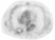
Transaxial PET image of lower thorax of a lesion the right lower lobe of the lung.
View main image(pt) in a separate viewing box
View second image(ct). CT of the chest at the same level as the right lower lobe lesion on PET.
View third image(pt). Transaxial PET image of the chest in the right upper lobe.
View fourth image(ct). CT of the chest at the level of the right upper lobe lesion.
Full history/Diagnosis is also available
Return to the Teaching File home page.