Case Author(s): Yungao Ding, M.D., Ph.D. and Jerold Wallis, M.D. , 12/05/2000 . Rating: #D., #Q.
Diagnosis: Lung cancer and pulmonary hamartoma
Brief history:
63 year old male with newly diagnosed right lung masses on CT of the chest
Images:

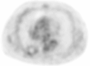
Transaxial PET image of lower thorax of a lesion the right lower lobe of the lung.
View main image(pt) in a separate image viewer
View second image(ct).
CT of the chest at the same level as the right lower lobe lesion on PET.
View third image(pt).
Transaxial PET image of the chest in the right upper lobe.
View fourth image(ct).
CT of the chest at the level of the right upper lobe lesion.
Full history/Diagnosis is available below
Diagnosis: Lung cancer and pulmonary hamartoma
Full history:
63 year old man presented with solitary brain mass and a right upper lobe lung mass. Both the brain mass and the right upper lung mass were proved to be squamous cell carcinoma upon biopsy, consistent with right upper lobe lung cancer with solitary brain metastasis. Chest X-ray and CT also show a densely calcified 3 cm right lower lobe mass consistent with a pulmonary hamartoma. Patient is evaluated with PET for additional body metastasis.
Radiopharmaceutical:
15.0 mCi F-18 FDG i.v.
Findings:
PET images of the body shows a photopenic focus with a rim of minimal FDG uptake in the right lower lobe. CT of the chest shows a densely calcified 3 cm mass with irregular margin in the posterior right lower lobe corresponding to the photopenic focus on PET imaging. The CT findings of a densely calcified lung mass on CT of the chest are consistent with a pulmonary hamartoma.
The soft tissue mass in the right upper lobe shows marked FDG uptake in the periphery of the lesion consistent with malignancy. The relatively photopenic center of the lesion corresponds to the low-attenuation center of the lung mass on CT consistent with central necrosis of the carcinoma.
Discussion:
Pulmonary hamartoma is the most common benign lung tumor found in 0.25% of general population and accounts for 6-8% of all solitary pulmonary lesions. The patient is usually asymptomatic. About 15% of the pulmonary hamartomas are calcified (almost pathognomonic if of chondroid "popcorn" type as in this case).
Hamartoma is composed of "normal" tissue with abnormal quantity and arrangement, and the glucose metabolism of the pulmonary hamartomas is therefore expected to be similar to the normal tissue. The photopenic center of the hamartoma in this case is likely due to the relative lack of cellularity in the central densely calcified portion of the lesion. The minimal uptake of FDG in the rim of the lesion is probably due to the relatively cellular uncalcified portion of the hamartoma and/or the surrounding compressed normal lung parenchyma.
Major teaching point(s):
Malignant lesions usually have increased anaerobic metabolism and significantly increased uptake of F-18 FDG while benign lesions and some slow-growing/low grade malignancy shows no dramatic increase of FDG uptake.
Differential Diagnosis List
Pulmonary hamartoma
Calcified granuloma
ACR Codes and Keywords:
References and General Discussion of PET Tumor Imaging Studies (Anatomic field:Lung, Mediastinum, and Pleura, Category:Neoplasm, Neoplastic-like condition)
Search for similar cases.
Edit this case
Add comments about this case
Return to the Teaching File home page.
Case number: pt037
Copyright by Wash U MO

