After viewing the image(s), the Full history/Diagnosis is available by using the link here or at the bottom of this page

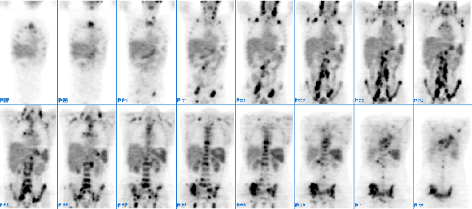
Coronal images from an F-18 FDG PET scan are shown.
View main image(pt) in a separate viewing box
View second image(pt). Selected coronal, axial, and sagittal views are shown
View third image(ct). Transaxial slice from CT scan through the pelvis is shown
View fourth image(ct). Another CT slice through the pelvis is shown.
Full history/Diagnosis is also available
Return to the Teaching File home page.