Case Author(s): Jeff Chesnut, D.O. and Farrokh Dedashti, M.D. , 6/9/99 . Rating: #D1, #Q5
Diagnosis: Hodgkin's Lymphoma
Brief history:
40 year old male.
Images:

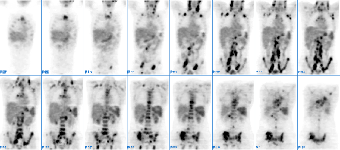
Coronal images from an F-18 FDG PET scan are shown.
View main image(pt) in a separate image viewer
View second image(pt).
Selected coronal, axial, and sagittal views are shown
View third image(ct).
Transaxial slice from CT scan through the pelvis is shown
View fourth image(ct).
Another CT slice through the pelvis is shown.
Full history/Diagnosis is available below
Diagnosis: Hodgkin's Lymphoma
Full history:
40 year old male with nodular sclerosing Hodgkin's lymphoma. PET study was
requested for staging.
Radiopharmaceutical:
F-18 Flourodeoxyglucose (FDG)
Findings:
There is extensive, intense increased metabolic activity in several nodal
chains including the anterior and posterior cervical chains, bilateral
supraclavicular nodes, bilateral axillary nodes, paratracheal, bilateral
hilar, retroperitoneal, iliac chain, and bilateral inguinal nodes. There is
also focal activity consistent with nodal disease involving the splenic
hilum, mesenteric, and peripancreatic region, as well as the gastrohepatic
ligament. These all correspond with enlarged nodes seen on outside CT, two
slices of which are reproduced here.
There are several foci of increased activity involving the ribs and
costovertebral junctions at several levels suggesting bone involvement.
There is also heterogenous increased activity involving the spine, most
prominent at S1, L4 and L5, and T9 through T11. There is also bone marrow
involvement of the entire pelvis, most prominent in the right iliac wing and the
region of the right sacroiliac joint. There is also increased activity
in the proximal right femur. There is diffuse, heterogenously increased
F-18 FDG activity in the entire spleen.
Discussion:
FDG-PET has a very high sensitivity (62% to 100%) in the detection
and staging of lymphoma, both Hodgkin's disease and Non-Hodgkin's. At our
institution, FDG-PET is rapidly supplanting/replacing Ga-67 scintigraphy as the
staging method of choice in lymphoma. Whereas about half of lymphomas are
gallium avid, nearly all lymphomas accumulate FDG. In addition, FDG-PET has been shown to be highly accurate in differentiating recurrent/residual tumor from scar. There is also evidence that FDG uptake shortly after institution of therapy is predictive of response.
In the first half of 1999, the Health Care Financing Administration agreed
to reimburse F-18 FDG PET for the staging of Hodgkin's and non-Hodgkin's
lymphoma.
Reference:
1. Clinical Positron Imaging 1998; 1:101-110.
Followup:
The patient received high dose chemotherapy.
Major teaching point(s):
1. F-18 FDG is very sensitive and specific for the staging of both Hodgkin's
and non-Hodgkin's lymphoma.
2. While only about half of all lymphomas are gallium avid, nearly all
lymphomas accumulate F-18 FDG.
ACR Codes and Keywords:
References and General Discussion of PET Tumor Imaging Studies (Anatomic field:Vascular and Lymphatic Systems, Category:Neoplasm, Neoplastic-like condition)
Search for similar cases.
Edit this case
Add comments about this case
Return to the Teaching File home page.
Case number: pt023
Copyright by Wash U MO

