After viewing the image(s), the Full history/Diagnosis is available by using the link here or at the bottom of this page
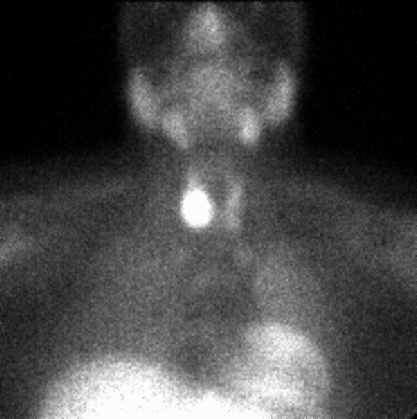
10 minute image from Tc-99m sestamibi parathyroid scintigram
View main image(pa) in a separate viewing box
View second image(pa). 2 hour delayed image from Tc-99m sestamibi parathyroid scintigram
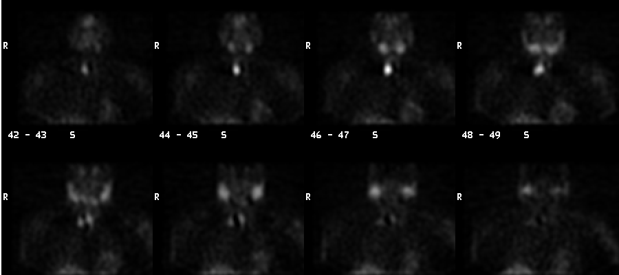
View third image(pa). Coronal SPECT images from Tc-99m sestamibi parathyroid scintigram, going from anterior to posterior.
Full history/Diagnosis is also available
Return to the Teaching File home page.