Case Author(s): Jeff Chesnut, D.O. and Jerold Wallis, M.D. , 6/9/99 . Rating: #D2, #Q4
Diagnosis: Thyroid adenoma
Brief history:
35 year old female with a palpable right neck nodule and elevated calcium.
Images:
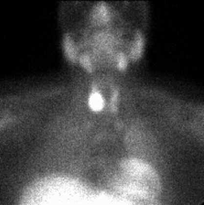
10 minute image from Tc-99m sestamibi parathyroid scintigram
View main image(pa) in a separate image viewer
View second image(pa).
2 hour delayed image from Tc-99m sestamibi parathyroid scintigram
View third image(pa).
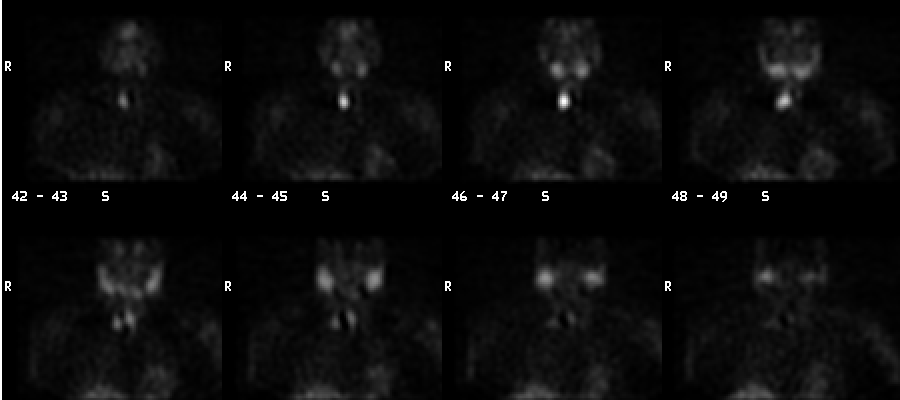
Coronal SPECT images from Tc-99m sestamibi parathyroid scintigram,
going from anterior to posterior.
Full history/Diagnosis is available below
Diagnosis: Thyroid adenoma
Radiopharmaceutical:
Tc-99m sestamibi
Findings:
There is a large nodule with intense increased uptake of Tc-99m sestamibi
projecting over the lower pole of the right lobe of the thyroid on the 10
minute image which persists on the 2 hr. image. Early SPECT images
demonstrate the position of the nodule to be anterior to the plane of the
right lobe of the thyroid, rather than along the posterior aspect
of the thyroid (as is more typical for parathyroid adenomas).
Discussion:
Tc-99m sestamibi is taken up by both normal thyroid and parathyroid tissue.
and should rapidly wash out. Both thyroid and parathyroid adenomas, however,
commonly have delayed wash-out of Tc-99m sestamibi. Thyroid adenoma is a well known
cause of false positive scintigrams when searching for parathyroid adenoma.
In this case, the anterior position of the nodule as well as the large size
suggest thyroid, rather than parathyroid, etiology.
The SPECT imaging is usually performed early so that sestamibi is still present
within the thyroid to provide landmarks.
Followup:
Excisional biopsy revealed this to be a large thyroid adenoma.
No additional foci suggestive of parathyroid adenomas were identified
on the scintigraphic study.
Major teaching point(s):
1. Both thyroid adenomas and parathyroid adenomas can have delayed wash-out
of Tc-99m sestamibi.
2. If SPECT scans are to be performed, they should be performed early so
that sestamibi in normal thyroid may be utilized for landmarks.
3. Large (palpable) size and anterior location both suggest thyroid adenoma rather
than parathyroid adenoma.
Differential Diagnosis List
1. Thyroid adenoma
2. Parathyroid adenoma
ACR Codes and Keywords:
References and General Discussion of Parathyroid Scintigraphy (Anatomic field:Face, Mastoids, and Neck, Category:Metabolic, endocrine, toxic)
Search for similar cases.
Edit this case
Add comments about this case
Return to the Teaching File home page.
Case number: pa006
Copyright by Wash U MO