back to index of exported cases

Unknown Case
Authored By: Farrokh Dehdashti and Xiaoni Hong.
Patient: 57 year old male
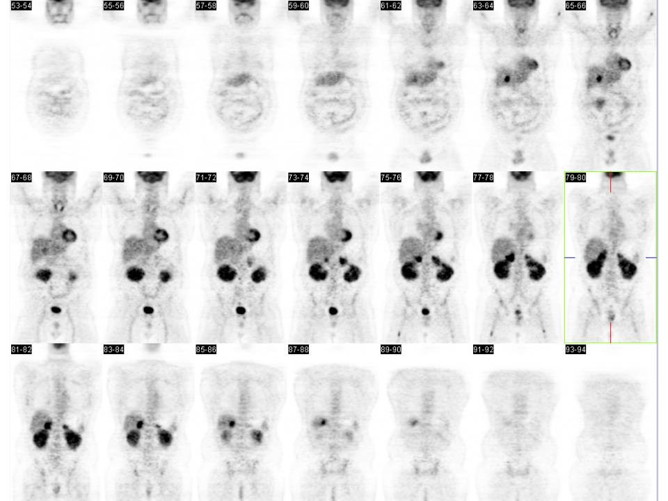
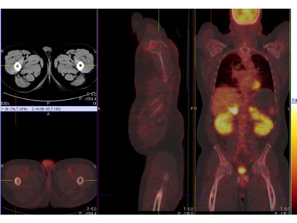
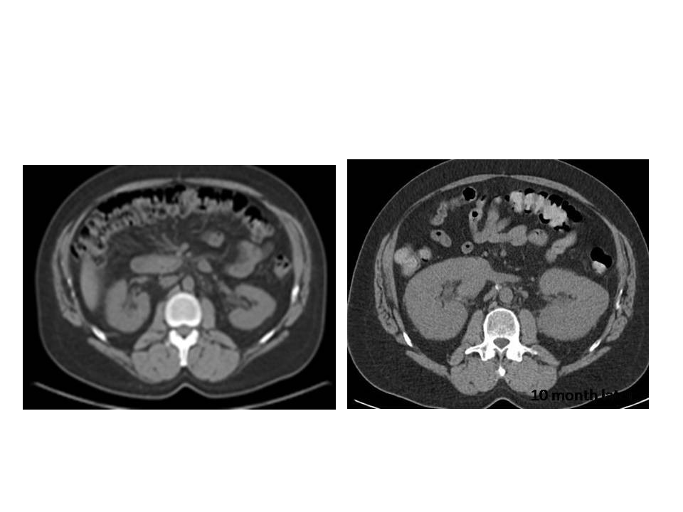
History: 56-year-old man with history of large B-cell non-Hodgkin's lymphoma, originally diagnosed three years prior to images shown here and subsequently treated with chemotherapy.
Image Size:[small][as-submitted]

Fig. 1
whole body follow up PET coronal images showed markedly FDG uptake in the kidenys and adrenal glands.

Fig. 2
Axial, sagittal, coronal fusion PET/CT images and CT axial image showed markedly FDG uptake in the kidenys and adrenal glands.

Fig. 3
compare axial non-contrast CT (left) from two months later with the original axial non-contrast CT (right) which shows enlarged kidneys.

Fig. 4
2 months later after chemotherapy the follow-up whole body PET coronal images reveal Minimal residual active disease in the right proximal femoral bone marrow and renal parenchyma.
Image Size:[small][as-submitted]

Comments:
No comments posted.
Additional Details:

Case [View Case with Diagnosis]Owner(s): Farrokh Dehdashti and Xiaoni HongLast Updated: 12-07-2011
Anatomy: Other   Pathology: Neoplasm
Modality: PETAccess Level: Readable by all users, writable by NucMed Certifiers
Keywords: ptnm

Case has been viewed 68 times.
Certified by Farrokh Dehdashti on 07-28-2010

The reader is fully responsible for confirming the accuracy of this content.
Text and images may be copyrighted by the case author or institution.