Return to Case List with Diagnoses or Case List as Unknowns

RECURRENT LYMPHOMA
Authored By: Farrokh Dehdashti and Xiaoni Hong.
Patient: 57 year old male
History: 56-year-old man with history of large B-cell non-Hodgkin's lymphoma, originally diagnosed three years prior to images shown here and subsequently treated with chemotherapy.
Image Size:[small][as-submitted]

Fig. 1
whole body follow up PET coronal images showed markedly FDG uptake in the kidenys and adrenal glands.

Fig. 2
Axial, sagittal, coronal fusion PET/CT images and CT axial image showed markedly FDG uptake in the kidenys and adrenal glands.

Fig. 3
compare axial non-contrast CT (left) from two months later with the original axial non-contrast CT (right) which shows enlarged kidneys.

Fig. 4
2 months later after chemotherapy the follow-up whole body PET coronal images reveal Minimal residual active disease in the right proximal femoral bone marrow and renal parenchyma.
Image Size:[small][as-submitted]

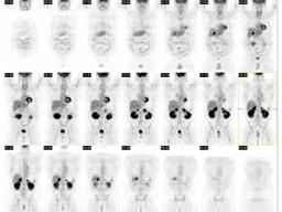
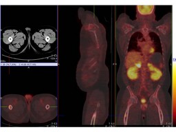
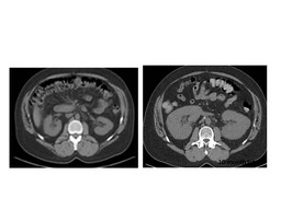
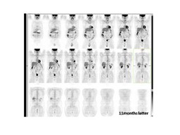
Findings:
FDG-PET/CT : The images demonstrated that the adrenal glands and kidneys are enlarged and show intense FDG uptake, highly suspicious for lymphoma involvement. Additionally, there is increased FDG uptake in an aortocaval lymph node at the level of renal vessels, suspicious for lymphoma recurrence. Focally intense FDG accumulation within the bone marrow of the left proximal humerus and proximal femoral shaft. These are also suspicious for lymphomatous involvement.  

Follow-up PET/CT after therapy (2 months later): Minimal residual active disease in the right proximal femoral bone marrow and renal parenchyma.
Diagnosis: Extranodal Lymphoma involing kidenys and adrenal glands.
General Discussion: Long History:56-year-old man with history of large B-cell non-Hodgkin's lymphoma, originally diagnosed 3 years ago and subsequently treated with chemotherapy. He experienced a recurrent retroperitoneal mass 15 months later. The patient was subsequently treated with further chemotherapy and underwent autologous stem cell transplant about one year ago. PET/CT study is requested for recurrence as the patient has been experiencing general malaise.

Radiopharmaceutical: 14.8 mCi F-18 Fluorodeoxyglucose (FDG) i.v.

Discussion: Extranodal spread of lymphoma often affects the genitourinary system, with the kidneys being the most commonly involved organs. Contrast–enhanced CT often used for the detection, diagnosis, staging, and response monitoring for renal lymphoma. MR imaging is particularly useful in patients in whom intravenous administration of iodinated contrast material is contraindicated. Ultrasonography (US), although very valuable for diagnosing lymphoma in the testis or epididymis, is less sensitive than CT and MR imaging for detecting renal lymphoma. Typical imaging findings of renal lymphoma include multiple poorly enhancing or hypoechoic masses, retroperitoneal tumors directly invading the kidneys, bilateral renal enlargement, and perirenal soft-tissue masses. FDG-PET has been shown to be an important technique for both staging and follow-up of nodal and extranodal lymphoma. PET–CT systems, which enable the performance of PET and CT data acquisition at the same setting without changing the patient's positioning, have been recently introduced in clinical practice. Lesions are characterized on the fused PET–CT images by both their metabolic status and their anatomic details. In general image fusion can also assist in the differentiation of physiologic and tumoral sites of FDG uptake.

References: 1. Sheila Sheth, MD, Syed Ali, MD and Elliot Fishman, MD .Imaging of Renal Lymphoma: Patterns of Disease with Pathologic Correlation. RadioGraphics 2006;26:1151-1168
2. Ur Metser, Odelia Goor, Hedva Lerman, Elizabeth Naparstek and Einat Even-Sapir.PET–CT of Extranodal Lymphoma.AJR 2004; 182:1579-1586

2.
  Even-Sapir E, Lievshitz G, Perry C, Herishanu Y, Lerman H, Metser U.  Fluorine-18 fluorodeoxyglucose PET/CT patterns of extranodal involvement in patients with Non-Hodgkin lymphoma and Hodgkin's disease.  Radiol Clin North Am. 2007 Jul;45(4):697-709.


Comments:
No comments posted.
Additional Details:

Case Number: 206900Owner(s): Farrokh Dehdashti and Xiaoni HongLast Updated: 12-07-2011
Anatomy: Other   Pathology: Neoplasm
Modality: PETAccess Level: Readable by all users, writable by NucMed Certifiers
Keywords: ptnm

Case has been viewed 68 times.
Certified by Farrokh Dehdashti on 07-28-2010

The reader is fully responsible for confirming the accuracy of this content.
Text and images may be copyrighted by the case author or institution.