Image Size: [small] [small] [as-submitted] [as-submitted]

 |
| Fig. 1 |
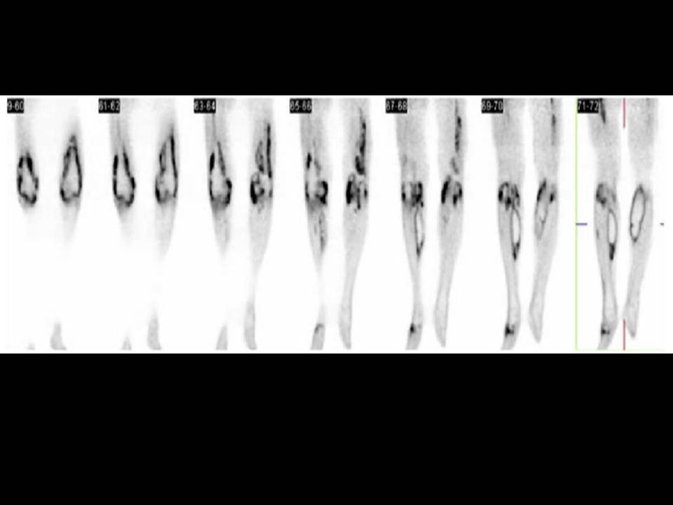
| Coronal PET images of the lower extremities reveal intense FDG uptake in the knee joints and various muscles in a pattern most compatible with infection. |
|

 |
| Fig. 2 |
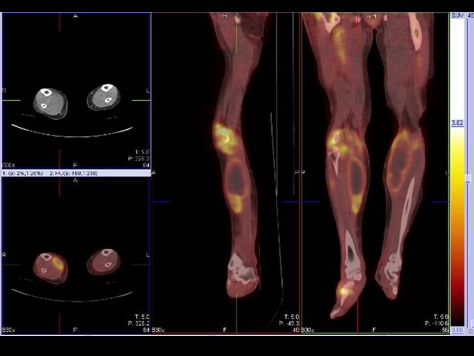
| PET/CT images of the lower extremities reveal large necrotic-appearing masses within each of the gastrocnemius muscles. Each is about 5 cm in diameter and measures 15 Hounsfield units, surrounded by a rim of marked FDG avidity. These are both most compatible with gastrocnemius abscesses or focal muscle liquefaction. Further FDG uptake is seen along the course of the right anterior tibialis muscle. There is a focus of marked FDG avidity in the right mid-foot and at the lateral aspect of the left forefoot.The right gluteal musculature is grossly enlarged with indistinct margins, also demonstrating moderate to marked FDG avidity. There is moderate uptake of FDG within the right hamstring muscle group. Marked FDG uptake is observed in a synovial distribution surrounding both knees. |
|

 |
| Fig. 3 |
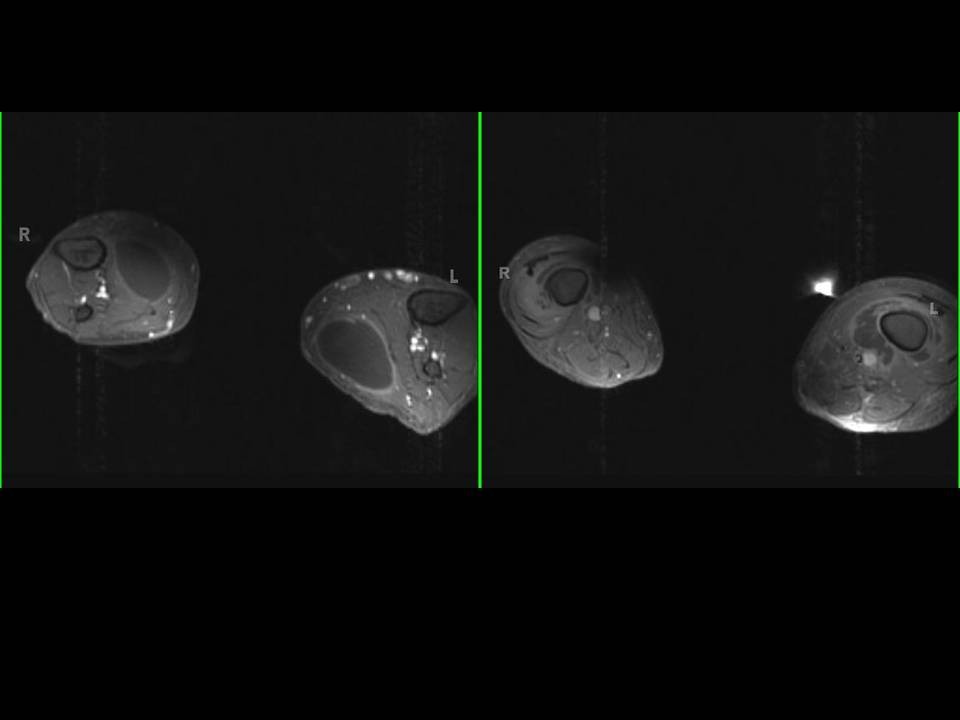
| Axial MRI T1 constrast of Lower extrimities reveal bilateral, moderate to large, fluid collections in both gastrocnemius muscles with smooth peripheral rim enhancement after contrast administration. |
|
Image Size: [small] [small] [as-submitted] [as-submitted]
|