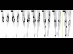
back to index of exported cases

Unknown Case
Authored By: Farrokh Dehdashti and Xiaoni Hong.
Patient: 55 year old female
History: This is a 55-year-old woman who was in her usual state of good health until approximately 1-1/2 weeks ago when she developed fever, back pain, and symptoms of malaise and an upper respiratory infection.
Image Size:[small][as-submitted]

Fig. 1
Coronal PET images of the lower extremities reveal intense FDG uptake in the knee joints and various muscles in a pattern most compatible with infection.
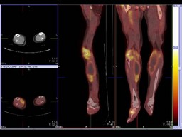
Fig. 2
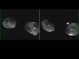
PET/CT images of the lower extremities reveal large necrotic-appearing masses within each of the gastrocnemius muscles. Each is about 5 cm in diameter and measures 15 Hounsfield units, surrounded by a rim of marked FDG avidity. These are both most compatible with gastrocnemius abscesses or focal muscle liquefaction. Further FDG uptake is seen along the course of the right anterior tibialis muscle. There is a focus of marked FDG avidity in the right mid-foot and at the lateral aspect of the left forefoot.The right gluteal musculature is grossly enlarged with indistinct margins, also demonstrating moderate to marked FDG avidity. There is moderate uptake of FDG within the right hamstring muscle group. Marked FDG uptake is observed in a synovial distribution surrounding both knees.

Fig. 3
Axial MRI T1 constrast of Lower extrimities reveal bilateral, moderate to large, fluid collections in both gastrocnemius muscles with smooth peripheral rim enhancement after contrast administration.
Image Size:[small][as-submitted]

Comments:
No comments posted.
Additional Details:

Case [View Case with Diagnosis]Owner(s): Farrokh Dehdashti and Xiaoni HongLast Updated: 12-07-2011
Anatomy: Skeletal System   Pathology: Infection
Modality: PETAccess Level: Readable by all users, writable by NucMed Certifiers
Keywords: ptnmACR: 40000.61400

Case has been viewed 53 times.
Certified by Farrokh Dehdashti on 07-07-2008

The reader is fully responsible for confirming the accuracy of this content.
Text and images may be copyrighted by the case author or institution.