back to index of exported cases

Unknown Case
Authored By: Keith Fischer and Chad Wright.
Patient: 66 year old male
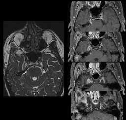
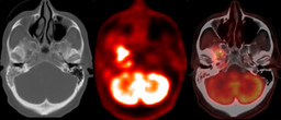
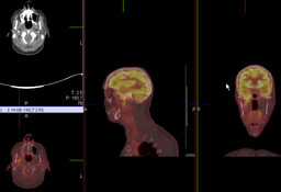
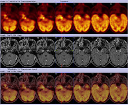
History: 66-year-old man with right parotid gland squamous cell carcinoma status post radiation who presents with double vision. Recent MRI suggested perineural spread of tumor along the right V3 nerve.
Image Size:[small][as-submitted]

Fig. 1
Contrast-enhanced MRI - T2 weighted axial image (left) and multiple serial post-contrast T1 weighted axial images (right)

Fig. 2
18F-FDG PET/CT - Axial CT (left), PET (middle), and fused PET/CT (right) images at the level of the skull base.

Fig. 3
18F-FDG PET/CT - Axial, sagittal and coronal fused PET/CT images demonstrating abnormally increased FDG uptake along the course of the right V3 nerve.

Fig. 4
18F-FDG PET fused with prior constrast-enhanced MRI - Axial PET (top), axial post-contrast T1 weighted MRI (middle), and fused PET/MRI (bottom) images at the level of the skull base.
Image Size:[small][as-submitted]

Comments:
No comments posted.
Additional Details:

Case [View Case with Diagnosis]Owner(s): Keith Fischer and Chad WrightLast Updated: 02-07-2013
Anatomy: Cranium and Contents   Pathology: Neoplasm
Modality: CT, MR, PETAccess Level: Readable and writable by Nuclear Medicine only

Case has been viewed 12 times.
Certified by Keith Fischer on 01-23-2013

The reader is fully responsible for confirming the accuracy of this content.
Text and images may be copyrighted by the case author or institution.