|
| Patient: 66 year old male |
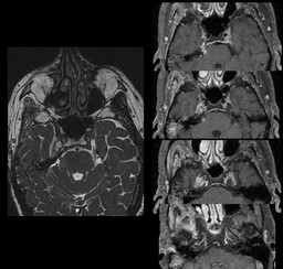
| History: 66-year-old man with right parotid gland squamous cell carcinoma status post radiation who presents with double vision. Recent MRI suggested perineural spread of tumor along the right V3 nerve. |
Image Size:
|
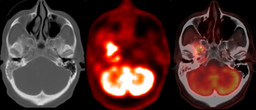
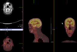
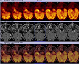
| Findings: Contrast-enhanced MRI: Ā Postsurgical changes consistent with a right parotidectomy are noted without evidence of enhancing tissue within the resection bed.Ā There is enhancement along the entire course of the right V3 nerve extending from the right pons, right trigeminal ganglion, right Meckel's cave, extending through the right foramen ovale, and into the right muscles of mastication.Ā Ā 18F-FDG PET/CT: Ā There is increased radiotracer uptake in the medial aspect of the right temporal lobe in the region of Meckel's cave and foramen ovale which extends to the anterior and right-sided pons.Ā There is also prominence of the right foramen ovale when compared with theĀleft.Ā Incidentally, there is diffuse moderately increased radiotracer uptakeĀin the thyroid gland, which can be seen in Hashimoto's thyroiditis.Ā Ā The PET images were also fused with the patient's prior contrast-enhanced MRI for further anatomic correlation and assessment. |
| Diagnosis: Perineural spread of recurrent right parotid squamous cell carcinoma along the right V3 nerve. |
| References: Fukui MB, Blodgett TM, Snyderman CH, Johnson JJ, Myers EN, Townsend DW and Meltzer CC.Ā Combined PET-CT in the Head and Neck.Ā Part 2.Ā Diagnostic Uses and Pitfalls of Oncologic Imaging.Ā Radiographics.Ā 2005.Ā Volume 25.Ā Pages 913-930. |
| Comments: No comments posted. |
| Additional Details:
Case Number: 387927 The reader is fully responsible for confirming the accuracy of this content. |