
 |
| Fig. 1 |
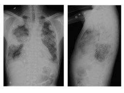
| PA and lateral chest radiography reveal multiple large bilateral pleural based masses with upper mediastinal lymphadenopathy and small bilateral pleural effusions. |
| 
 |
| Fig. 2 |
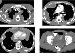
| Axial chest and pelvis contrast CT demonstrated heterogeneously enhancing nodules involving the greater portion of the pleural surfaces in both lungs, with extension into the pericardium and apparent extension into the chest wall. Ill-defined large mass around the rectosigmoid region that could represent a large occult adenocarcinoma of the rectum. |
|

 |
| Fig. 3 |
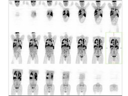
| coronal whole body PET images demonstrated intense FDG-avid extensive bilateral pleural-based soft tissue masses with extension into the right pulmonary parenchyma and along the pericardium, with an FDG-avid right axillary lymph node. No abnormal FDG uptake in CT described ill-defined large mass in the rectosigmoid region |
| 
 |
| Fig. 4 |
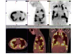
| Axial,sagittal,coronal PET and fusion images demonstrated intense FDG-avid extensive bilateral pleural-based soft tissue masses with extension into the right pulmonary parenchyma and along the pericardium, with an FDG-avid right axillary lymph node. |
|

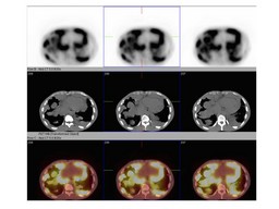 |
| Fig. 5 |
| Axial PET, CT and fusion images demonstrated intense FDG-avid extensive bilateral pleural-based soft tissue masses with extension into the right pulmonary parenchyma and along the pericardium. |
|