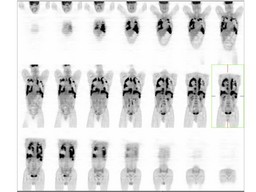
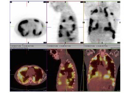
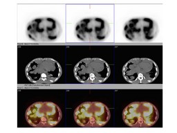
Return to Case List with Diagnoses or Case List as Unknowns

POORLY DIFFERENTIATED CARCINOMA
Authored By: Xiaoni Hong and Jerold Wallis, Assoc Prof of Radiology.
Patient: male
History: 31 old year old male  complains of  sharp chest pain, shortness of breath, cough, fever, night sweats and 15 lb weight loss.
Image Size:[small][as-submitted]

Fig. 1
PA and lateral chest radiography reveal multiple large bilateral pleural based masses with upper mediastinal lymphadenopathy and small bilateral pleural effusions.

Fig. 2
Axial chest and pelvis contrast CT demonstrated heterogeneously enhancing nodules involving the greater portion of the pleural surfaces in both lungs, with extension into the pericardium and apparent extension into the chest wall. Ill-defined large mass around the rectosigmoid region that could represent a large occult adenocarcinoma of the rectum.

Fig. 3
coronal whole body PET images demonstrated intense FDG-avid extensive bilateral pleural-based soft tissue masses with extension into the right pulmonary parenchyma and along the pericardium, with an FDG-avid right axillary lymph node. No abnormal FDG uptake in CT described ill-defined large mass in the rectosigmoid region

Fig. 4
Axial,sagittal,coronal PET and fusion images demonstrated intense FDG-avid extensive bilateral pleural-based soft tissue masses with extension into the right pulmonary parenchyma and along the pericardium, with an FDG-avid right axillary lymph node.

Fig. 5
Axial PET, CT and fusion images demonstrated intense FDG-avid extensive bilateral pleural-based soft tissue masses with extension into the right pulmonary parenchyma and along the pericardium.
Image Size:[small][as-submitted]

Findings:


FDG PET
: Intense FDG-avid extensive bilateral pleural-based soft tissue masses with extension into the right pulmonary parenchyma and along the pericardium, with an FDG-avid right axillary lymph node.No abnormal FDG uptake in previously described ill-defined large mass in the rectosigmoid region.No other sites of tumor visualized to suggest an extra-pulmonary location for the primary tumor.

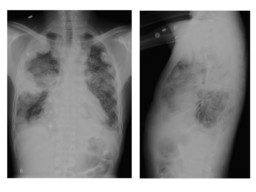
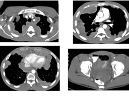
CT of chest : Diffuse, heterogeneously enhancing nodules involving the greater portion of the pleural surfaces in both lungs, with extension into the pericardium and apparent extension into the chest wall.Ill-defined large mass around the rectosigmoid region that could represent a large occult adenocarcinoma of the rectum.


Diagnosis: POORLY DIFFERENTIATED CARCINOMA
General Discussion:

Long history: 31 old year old male  complains of  sharp chest pain, shortness of breath, cough, fever, night sweats and 15 lb weight loss. He has  poorly differentiated adenocarcinoma of unknown primary origin as demonstrated on recent biopsy. PET/CT study is requested for evaluate for primary tumor.

Radiopharmaceutical: 14.9 mCi F-18 Fluorodeoxyglucose i.v.


Followup pathology

A poorly differentiated carcinoma. Patent underwent chemotherapy: Carboplatin, Taxol, Alimta 2 cycles.  Disease worsened without any response to therapy.

Comments:
No comments posted.
Additional Details:

Case Number: 206897Owner(s): Xiaoni Hong and Jerold Wallis, Assoc Prof of RadiologyLast Updated: 12-07-2011
Anatomy: Other   Pathology: Neoplasm
Modality: PETAccess Level: Readable by all users, writable by NucMed Certifiers
Keywords: ptnmACR: 60000.32000

Case has been viewed 31 times.
Certified by Jerold Wallis on 06-17-2009

The reader is fully responsible for confirming the accuracy of this content.
Text and images may be copyrighted by the case author or institution.