
 |
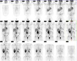
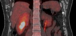
| Fig. 1 |
| Coronal FDG PET images demonstrate tracer avid tissue in the right posterior neck and right retroperitoneum. |
|
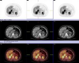 |
| Fig. 2 |
| FDG PET, CT, and fused PET/CT images demonstrate a tracer avid mass of the right kidney as well as enlarged, tracer avid lymph nodes. |
|
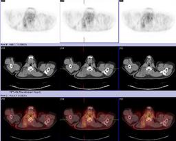 |
| Fig. 3 |
| FDG PET, CT, and fused PET/CT images demonstrate a mildly tracer avid mass in the soft tissues of the lower right neck. |
|
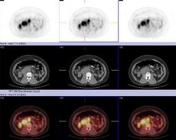 |
| Fig. 4 |
| FDG PET, CT, and fused PET/CT images demonstrated enlarge, tracer avid right retroperitoneal lymph nodes |
|
 |
| Fig. 5 |
| Fused coronal FDG PET and CT images demonstrate a moderately tracer avid soft tissue mass involving the upper pole of the right kidney. Intense FDG activity is seen in the right renal pelvis which is displaced inferiorly. |
|
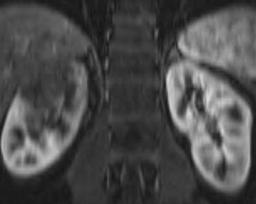 |
| Fig. 6 |
| Coronal gadolinium enhanced T1WI demonstrates an infiltrative soft tissue mass involving the upper pole of the right kidney. |
|
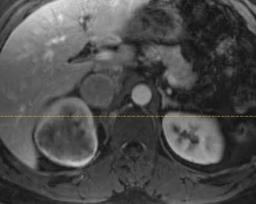 |
| Fig. 7 |
| Axial gadolinium enhanced T1WI demonstrates an infiltrating soft tissue mass centered in the renal medulla enhancing less than the surrounding renal cortex. |
|