Return to Case List with Diagnoses or Case List as Unknowns

MEDULLARY CARCINOMA OF THE KIDNEY
Authored By: Delphine Chen and Brandon Peters.
Patient: 39 year old male
History: 40 year old male without significant prior medical history was found to have enlarged retroperitoneal lymph nodes and an enlarging neck mass. 
Image Size:[small][as-submitted]

Fig. 1
Coronal FDG PET images demonstrate tracer avid tissue in the right posterior neck and right retroperitoneum.

Fig. 2
FDG PET, CT, and fused PET/CT images demonstrate a tracer avid mass of the right kidney as well as enlarged, tracer avid lymph nodes.

Fig. 3
FDG PET, CT, and fused PET/CT images demonstrate a mildly tracer avid mass in the soft tissues of the lower right neck.

Fig. 4
FDG PET, CT, and fused PET/CT images demonstrated enlarge, tracer avid right retroperitoneal lymph nodes

Fig. 5
Fused coronal FDG PET and CT images demonstrate a moderately tracer avid soft tissue mass involving the upper pole of the right kidney. Intense FDG activity is seen in the right renal pelvis which is displaced inferiorly.

Fig. 6
Coronal gadolinium enhanced T1WI demonstrates an infiltrative soft tissue mass involving the upper pole of the right kidney.

Fig. 7
Axial gadolinium enhanced T1WI demonstrates an infiltrating soft tissue mass centered in the renal medulla enhancing less than the surrounding renal cortex.
Image Size:[small][as-submitted]

Findings:

Radiopharmaceutical:  14.7 mCi 18-Flourodeoxyglucose.
 
FDG PET/CT of the body demonstrates enlarged and tracer avid lymph nodes in the right retroperitoneum and around the inferior vena cava.  Additionally, there is a mildly tracer avid soft tissue mass in the muculature of the right lower neck/shoulder.  An infiltrative tracer avid mass is demonstrated centered in the hilum of the upper pole of the right kidney.  Gadolinium enhanced MRI demonstrates the soft tissue mass centered in the hilum of the upper pole of the right kidney.  The mass enhances signficantly less than the surrounding relatively preserved renal cortex.

The differential diagnosis includes lymphoma, atypical renal cell carcinoma, transitional cell carcinoma, or other less common neoplasms of the renal pelvis such as collecting duct carcinoma, rhabdoid tumor, or renal medullary carcinoma. 

Diagnosis: Renal medullary carcinoma
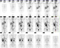
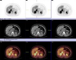
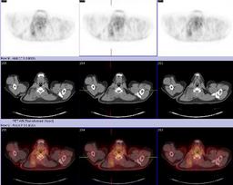
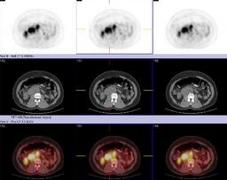
General Discussion:

40 year old male with no known medical history was found to have enlarged retrocrural lymph nodes which were subsequently biopsied and found to be poorly differentiated carcinoma.   Based on the location and appearance of the lesion, the patient was tested for sickle trait and found to be positive.  Review of the original lymph node pathology led to a presumptive diagnosis of medullary carcinoma of the kidney.

In 1995, Davis et al (1) first described renal medullary carcinoma as the seventh nephropathy of sickle cell disease.  The cancer is usually large and metatstatic by the time of detection.  It often presents with flank pain and hematuria.  The disease has a strong association with sickle cell trait but not with sickle cell disease.   Various cytogenetic abnormalities have been found in renal medullary carcinoma, including abnormalities of chromosome 11p, where the beta-globin gene is located.   However, no definitive direct chromosomal or environmental link has been established as the cause of this disease.  Like in this patient, several studies have shown renal medullary carcinoma has a striking tendancy to be right sided.  The most common sites of metastasis are the lymph nodes, lungs, liver, and peritoneum.  Many treatments have been tried including surgery, various chemotherapies, and radiation.  However, the prognosis remains dismal with medial survival four months from diagnosis (2).

References:

1.  Davis CJ Jr et al.  Renal Medullary Carcinoma.  The seventh sickle cell nephropathy.  Am J Surg Pathol 1995; 19: 1-11


2.  Noguera-Irizarry et al.  Renal Medullary Carcinoma:  Case report and review of the literature.  Am J Clin Onc 2003; Oct. 26(5) 489-492.

Comments:
No comments posted.
Additional Details:

Case Number: 206895Owner(s): Delphine Chen and Brandon PetersLast Updated: 12-07-2011
Anatomy: Genitourinary (GU)   Pathology: Neoplasm
Modality: CT, MR, PETAccess Level: Readable by all users, writable by NucMed Certifiers
Keywords: ptnmACR: 80000.32000

Case has been viewed 62 times.
Certified by Delphine Chen on 01-30-2009

The reader is fully responsible for confirming the accuracy of this content.
Text and images may be copyrighted by the case author or institution.