 |
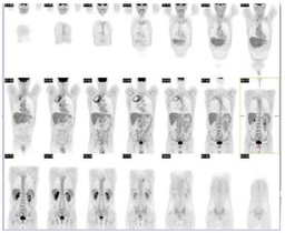
| Fig. 1 |
| Coronal PET images reveal a 1. 9 x 8.5 x 6.1 cm cavitary lesion in the apical segment of the right upper lobe demonstrating increased FDG uptake along its periphery. There are several associated subcentimeter pulmonary nodules in both right upper and right middle lobes, as well as tree-and-bud-like infiltrate in the right middle lobe. There is also markedly increased FDG uptake in a normal-sized right hilar lymph node. |
|
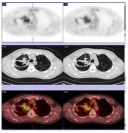 |
| Fig. 2 |
| Axial PET/CT images reveal a 1. 9 x 8.5 x 6.1 cm cavitary lesion in the apical segment of the right upper lobe demonstrating increased FDG uptake along its periphery. There are several associated subcentimeter pulmonary nodules in both right upper and right middle lobes, as well as tree-and-bud-like infiltrate in the right middle lobe. There is also markedly increased FDG uptake in a normal-sized right hilar lymph node. |
|