Return to Case List with Diagnoses or Case List as Unknowns

INVASIVE PULMONARY ASPERGILLOSIS
Authored By: Xiaoni Hong and Jerold Wallis, Assoc Prof of Radiology.
Patient: 57 year old male
History: 57 y.o. male with progressive cough.
Image Size:[small][as-submitted]

Fig. 1
Coronal PET images reveal a 1. 9 x 8.5 x 6.1 cm cavitary lesion in the apical segment of the right upper lobe demonstrating increased FDG uptake along its periphery. There are several associated subcentimeter pulmonary nodules in both right upper and right middle lobes, as well as tree-and-bud-like infiltrate in the right middle lobe. There is also markedly increased FDG uptake in a normal-sized right hilar lymph node.

Fig. 2
Axial PET/CT images reveal a 1. 9 x 8.5 x 6.1 cm cavitary lesion in the apical segment of the right upper lobe demonstrating increased FDG uptake along its periphery. There are several associated subcentimeter pulmonary nodules in both right upper and right middle lobes, as well as tree-and-bud-like infiltrate in the right middle lobe. There is also markedly increased FDG uptake in a normal-sized right hilar lymph node.

Fig. 3
Chest radiography reveal a large cavitary mass in the right lung apex that measures approximately 8 cm in diameter. There is thickening of the wall of this lesion superiorly and medially.
Image Size:[small][as-submitted]

Findings:

Long history: 57 y.o. heavy smoker male with progressive cough , who has cavitary right upper lung mass seen on the outside hospital CT scan. The study was requested to evaluate for malignancy and for staging.

Radiopharmaceutical: 18.3 mCi F-18 Fluorodeoxyglucose i.v.

Findings: 

FDG PET:1. there is a 1. 9 x 8.5 x 6.1 cm cavitary lesion in the apical segment of the right upper lobe demonstrating increased FDG uptake along its periphery. There are several associated subcentimeter pulmonary nodules in both right upper and right middle lobes, as well as tree-and-bud-like infiltrate in the right middle lobe. There is also markedly increased FDG uptake in a normal-sized right hilar lymph node. Based on the FDG uptake, this could represent malignancy or infection. Based on the CT appearance, the findings are concerning for an infectious process such as tuberculosis or fungal infection. Less likely, this could represent necrotizing pneumonia or squamous cell carcinoma. Correlation with expectorated sputum and bronchoscopy findings is suggested.

2. Moderate degree emphysema with upper lobe predominance.


Chest X-Ray: There is a large cavitary mass in the right lung apex that measures approximately 8 cm in diameter. There is thickening of the wall of this lesion superiorly and medially.

Diagnosis: Invasive pulmonary aspergillosis
General Discussion:

Follow up:
Bronchoalveolar lavage of the right upper lobe  was positive for Aspergillus fungatus.


Discussion: MAI Complex:
Nontuberculous mycobacteria associated with lung disease (COPD) or immunocompromise.Clinical symptoms are like TB.No human to human spread.

Radiologic appearance in non-immunocompromised patients (usually COPD):
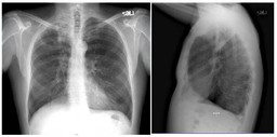
• CXR-linear and nodular opacities in apical and posterior upper lobe and superior lower lobe, bronchiectasis, fibrosis, atelectasis
• CT-centrilobular nodules (tree-in-bud), multilobe bronchiectasis (often lingula and RML), consolidation, cavitation, fibrosis, atelectasis

Radiologic appearance in immunocompromised patients can vary:
• Normal,  pleural effusion, consolidation, miliary

• Pulmonary Aspergillosis: Saprophytic aspergillosis (aspergilloma,mycetoma, fungus ball)
– Usually colonizes pre-existing cavity (TB, sarcoid, neoplasm)
– Intracavitary mass, air-crescent

• Allergic bronchopulmonary aspergillosis
– Allergic reaction to antigens
– Associated with asthma and CF
– Consolidation and bronchiectasis

• Chronic necrotizing pulmonary aspergillosis (semi-invasive)
– Associated with mild immunocompromise (steroids), DM, alcohol, COPD
– Upper lobe cavitary consolidation

• Invasive pulmonary aspergillosis
– Associated with neutropenia of AIDS, malignancy, transplant
– Early nodules and air-space consolidation, late cavitary lesions, hemorrhagic necrosis (halo sign)

Comments:
No comments posted.
Additional Details:

Case Number: 206889Owner(s): Xiaoni Hong and Jerold Wallis, Assoc Prof of RadiologyLast Updated: 12-07-2011
Anatomy: Other   Pathology: Infection
Modality: PETAccess Level: Readable by all users, writable by NucMed Certifiers
Keywords: ptnmACR: 60000.25000

Case has been viewed 54 times.
Certified by Jerold Wallis on 06-17-2009

The reader is fully responsible for confirming the accuracy of this content.
Text and images may be copyrighted by the case author or institution.