Return to Case List with Diagnoses or Case List as Unknowns

ESOPHAGEAL CANCER WITH RIB FRACTURES DUE TO CARDIOVASCULAR RESUSCITATION
Authored By: Farrokh Dehdashti and Peter Phan.
Patient: 50 year old
History: 50-year-old man with known malignancy and a complicated hospital course.
Image Size:[small][as-submitted]

Multimedia: 206886_1_submitted.avi
Initial Staging PET-CT

Multimedia: 206886_2_submitted.avi
Follow-up PET-CT

Fig. 3
Axial images from follow-up PET-CT

Fig. 4
Axial images from follow-up PET-CT

Fig. 5
Chest radiograph following the second PET-CT
Image Size:[small][as-submitted]

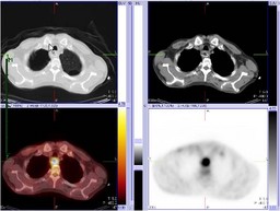
Findings:

Initial PET-CT:

RADIOPHARMACEUTICAL: 15 mCi F-18 Fluorodeoxyglucose i.v.

Two foci of markedly increased FDG uptake in the thickening of the mid esophagus, suspicious for malignancy.
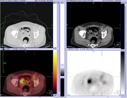

Follow up PET-CT:

RADIOPHARMACEUTICAL: 13.6 mCi F-18 Fluorodeoxyglucose i.v.

1. Two persistent foci of markedly increased FDG uptake involving the thickened mid esophagus, not significantly changed.


2. Multiple new rib and sternal fractures, likely related to cardiac resuscitation.


3. Diffuse mild increased FDG uptake in the left inguinal soft tissue, likely inflammatory in nature and related to vascular catheterization.

4. Free intraperitoneal air related to recent laproscopic procedure.

Diagnosis: Initial PET-CT: Esophageal cancer

Follow up PET-CT: Esophageal cancer with multiple rib and sternal fractures, related to cardiac resuscitation.
General Discussion: The patient is a 50-year-old man with esophageal cancer, status post esophageal stent. His first PET-CT for initial staging did not reveal any evidence of metastasis.  Subsequently, he was scheduled for an esophagectomy.  However, the patient had several episodes of hypotension during the induction of anesthesia and then coded, requiring cardiopulmonary resusitation. The surgery was aborted at that time.  After a long hospital course that included cardiovascular intervention (complicated by a left inguinal hematoma) and a laproscopic surgical procedure (free intraperitoneal air), the patient recovered.  Given the long time interval since his prior exam, the patient underwent a second PET-CT study for restaging. The main difference between the two studies were new areas of increased uptake within the sternum ans ribs.  The history and the pattern of FDG uptake are very important in order to avoid interpreting benign lesions such as fractures as metastases. 

The treatment of esophageal cancer is highly dependent on the initial stage.  The presence of local nodal metastasis may dictate the need for multimodality treatment beyond esophagectomy.  The accuracy of PET-CT for the detection of nodal disease is improved over CT alone, but remains overall low, requiring nodal sampling for patients who are surgical candidates.  The reported sensitivity and specificity for local nodal disease ranges from 22% to 71% and 73% to 100%, respectively [1].  The detection of distant metastatic disease, which precludes surgical intervention, is also more superior to other diagnostic imaging modalities.  PET-CT can also help with prognostic assessment, with one study showing a worse prognosis in patient with primary tumors that has SUV greater than 7, compared to those with SUV less than 3 [1].
References: [1] Dehdashti, Farrokh. PET and PET/CT in Esophageal and Gastric Cancers.  In: Valk P, ed. Positron Emission Tomography. London: Springer-Verlag; 2006: 165-171.
Comments:
No comments posted.
Additional Details:

Case Number: 206886Owner(s): Farrokh Dehdashti and Peter PhanLast Updated: 12-07-2011
Anatomy: Gastrointestinal (GI)   Pathology: Neoplasm
Modality: Conventional Radiograph, PETAccess Level: Readable by all users, writable by NucMed Certifiers
Keywords: ptnmACR: 70000.32000, 40000.41900

Case has been viewed 41 times.
Certified by Farrokh Dehdashti on 05-02-2008

The reader is fully responsible for confirming the accuracy of this content.
Text and images may be copyrighted by the case author or institution.