
 |
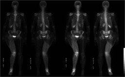
| Fig. 1 |
| Whole body bone scintigraphy |
|
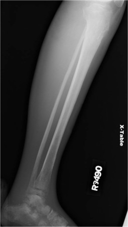 |
| Fig. 2 |
| Radiograph of the right lower extremity: Multifocal destructive bone lesions in the right distal femur, right tibia, and right distal fibula with severe soft tissue swelling of the right leg |
|
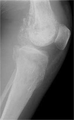 |
| Fig. 3 |
| Radiograph of the right lower extremity: Multifocal destructive bone lesions in the right distal femur, right tibia, and right distal fibula with severe soft tissue swelling of the right leg. |
|
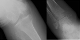 |
| Fig. 4 |
| Radiograph of the right lower extremity: Multifocal destructive bone lesions in the right distal femur, right tibia, and right distal fibula with severe soft tissue swelling of the right leg. |
|
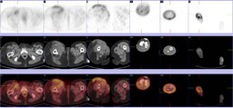 |
| Fig. 5 |
| PET/CT of the right lower extremity: An infiltrative process with soft tissue attenuation is seen in the entire right lower extremity with moderate to marked heterogeneous FDG uptake. |
|