Return to Case List with Diagnoses or Case List as Unknowns

EPITHELIOID ANGIOSARCOMA OF THE LEG
Authored By: Farrokh Dehdashti and Peter Phan.
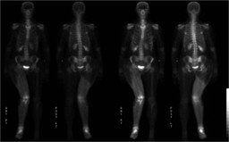
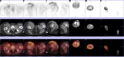
Patient: 50 year old
History: 50 year old female with leg pain.
Image Size:[small][as-submitted]

Fig. 1
Whole body bone scintigraphy

Fig. 2
Radiograph of the right lower extremity: Multifocal destructive bone lesions in the right distal femur, right tibia, and right distal fibula with severe soft tissue swelling of the right leg

Fig. 3
Radiograph of the right lower extremity: Multifocal destructive bone lesions in the right distal femur, right tibia, and right distal fibula with severe soft tissue swelling of the right leg.

Fig. 4
Radiograph of the right lower extremity: Multifocal destructive bone lesions in the right distal femur, right tibia, and right distal fibula with severe soft tissue swelling of the right leg.

Fig. 5
PET/CT of the right lower extremity: An infiltrative process with soft tissue attenuation is seen in the entire right lower extremity with moderate to marked heterogeneous FDG uptake.
Image Size:[small][as-submitted]

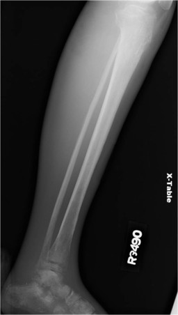
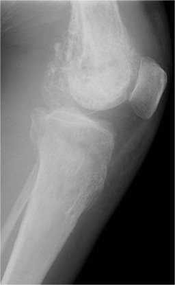
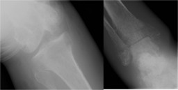
Findings: Bone scan with radiographic correlates: Diffuse increased uptake is noted throughout the soft tissues of the right leg. Focal increased uptake about the right knee is seen, involving the distal femur and proximal tibia.  This corresponds to a destructive process seen on the radiographs. Increased uptake in the right ankle is also present, probably related to severe osteoporosis.
Diagnosis: Epithelioid angiosarcoma of the right lower extremity
General Discussion: RADIOPHARMACEUTICAL: 21.9 mCi Tc-99m MDP i.v.

Based on the bone scan alone, the diffuse soft tissue uptake within the right leg is likely to represent chronic lymphedema from prior nodal dissection or venous obstruction. Electrical burns may have similar appearance.


However, further review of the patient's chart reveals that she had a soft tissue hemangioma resected over 20 years ago and that her leg swelling and skin induration has progressively worsened. Biopsy of the skin thickening revealed underlying malignancy: epithelioid angiosarcoma.


Cutaneous epithelioid angiosarcoma is a rare malignant vascular tumor that is difficult to diagnosis, often confused with a carcinoma or melanoma.  It presents predominately in middle to late adulthood males. Besides cutaneous in origin,  this malignancy has been document to arise from the the skeleton as well as visceral organs.  It has been shown that epithlioid angiosarcoma can arise from an irradiated hemangioma, but there is evidence that angiosarcomatous transformation from a benign nonirradiated vascular lesion is possible. The progression of epithelioid angiosarcoma is indolent, but some case reports suggest the presence of systemic metastasis within one year.

References: Rossi, Sabrina. "Angiosarcoma Arising in Hemangioma/Vascular Malformation: Report of Four Cases and Review of the Literature", The American Journal of Surgical Pathology. Volume 26(10), October 2002, pp 1319-1329.

Prescott, R.J..  "Cutaneous Epithelioid Angiosarcoma: A Clinicalpathological Study of Four cases", Histopathology 1994, 25, 421-429.  
Comments:
No comments posted.
Additional Details:

Case Number: 206885Owner(s): Farrokh Dehdashti and Peter PhanLast Updated: 12-07-2011
Anatomy: Skeletal System   Pathology: Neoplasm
Modality: Nuc Med, PETAccess Level: Readable by all users, writable by NucMed Certifiers
Keywords: bsnmACR: 40000.32400

Case has been viewed 28 times.
Certified by Farrokh Dehdashti on 07-08-2008

The reader is fully responsible for confirming the accuracy of this content.
Text and images may be copyrighted by the case author or institution.