| Additional Details:
Case [View Case with Diagnosis] Owner(s): Asif Moinuddin and Barry Siegel, Prof of RadiologyLast Updated: 12-07-2011 Owner(s): Asif Moinuddin and Barry Siegel, Prof of RadiologyLast Updated: 12-07-2011
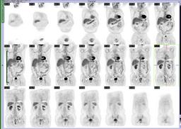
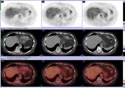
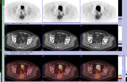
Anatomy: Spine and Peripheral Nervous System Pathology: Neoplasm
Modality: Nuc Med, PETAccess Level: Readable by all users, writable by NucMed Certifiers
Keywords: ptnm, chordoma, sarcoma, teratomaACR: 30000.32700
Case has been viewed 31 times.
Certified by Barry Siegel on 06-13-2009The reader is fully responsible for confirming the accuracy of this content.
Text and images may be copyrighted by the case author or institution.
|