|
| Patient: 61 year old male |
| History: 76-year-old man with esophageal adenocarcinoma post chemoradiation therapy and a sacrococcygeal chordoma. |
Image Size:
|
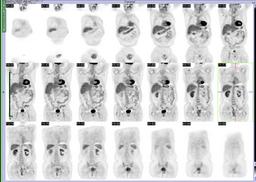
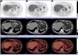
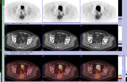
| Findings: RADIOPHARMACEUTICAL: 15.35 mCi F-18 Fluorodeoxyglucose i.v. FINDINGS: There is a focus of mildly increased FDG uptake in the distal esophagus that on the CT images correlates to a circumferentially thickened distal esophagus. The maximum standardized uptake value (SUV) is 4.8 in this focus. This could represent radiation changes, but residual tumor cannot be entirely excluded. A second focus of heterogeneous FDG uptake is seen in the sacrococcygeal region, representing the known chordoma. It correlates to a heterogeneous-attenuation pelvic mass that appears more dense centrally than peripherally. It measures 8.8 x 6.4 cm in its greatest dimension. The maximum SUV is 5.6. The increased FDG uptake is predominantly central and corresponds to the denser portion of the soft tissue component on the CT images. The mass is located within the lower presacral region and the precoccygeal space and displaces the rectum anteriorly. It appears to have destroyed the left side of the lower sacral segment and completely destroys the coccyx. It extends into soft tissues posterior to the plane of the sacrum and into the perianal region, particularly on the left side. There is increased FDG uptake in a right axillary nodal focus, most likely as a result of migration of tracer extravasated at the injection site. Post radiation changes involving the r thoracic and upper lumbar spine are also seen. |
| Diagnosis: Sacrococcygeal chordoma |
| General Discussion: FOLLOW-UP: The patient underwent surgical resection and the pathological examination demonstrated chordoma with sarcomatous features. |
| References: F-18 FDG PET/CT evaluation of sacrococcygeal chordoma. Clin Nucl Med. 2008 Dec;33(12):906-8. |
| Comments: No comments posted. |
| Additional Details:
Case Number: 206880 The reader is fully responsible for confirming the accuracy of this content. |