Return to Case List with Diagnoses or Case List as Unknowns

RECURRENT/ RESIDUAL BRAIN METASTATIC SMALL CELL CARCINOMA
Authored By: Xiaoni Hong and Akash Sharma.
Patient: 60 year old female
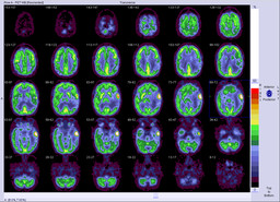
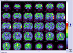
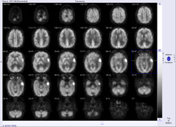
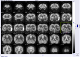
History:

61-year-old female with small cell lung cancer and prior intracranial metastatic disease.

Image Size:[small][as-submitted]

Fig. 1
Markedly hypermetabolic left temporal lobe lesion

Fig. 2
Markedly hypermetabolic left temporal lobe lesion

Fig. 3
Markedly hypermetabolic left temporal lobe lesion

Fig. 4
Markedly hypermetabolic left temporal lobe lesion

Fig. 5
Fused PET-MR demonstrates hypermetabolism corresponds to the enhancing mass

Fig. 6
Brain MRI T2W and postconterast reveal a left temporal lobe mass with maximal axial dimension of 4.1cm. Surrounding vasogenic edema. Mild midline shift to the right of 5mm.
Image Size:[small][as-submitted]

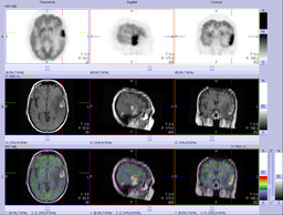
Findings:

FDG PET:  Highly FDG avid left temporal mass, with mild surround hypometabolism.  No other lesions.


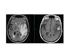
Brain MRI: Left temporal lobe mass with maximal axial dimension of 4.1cm, with surrounding vasogenic edema. Mild midline shift to the right of 5 mm.

DDx:

Recurrent or residual brain metastatic disease


Primary brain tumor


Infection/Herpes encephalitis


Lymphoma

Diagnosis: Recurrent or residual brain metastatic small cell carcinoma
General Discussion:

The patient is a 61-year-old female with small cell lung cancer and prior intracranial metastatic disease. She underwent whole brain radiation as well as gamma knife radiation in 2003. She had surgical resection of a cerebral lesion in August 2007.  A subsequent MRI revealed abnormal lesions suggestive of recurrent disease versus radiation necrosis. 

Radiopharmaceutical: 10 mCi F-18 Fluorodeoxyglucose i.v.

Follow up: Craniotomy/ brain tumor excision demonstrates recurrent/ residual brain metastatic small cell carcinoma. 

Teaching points:

  •  Brain PET/CT can be used for differentiating radiation necrosis verse residual tumor. Usually, radiation necrosis don’t have FDG uptake.


  • Brain PET/CT may also differentiate lymphoma verse toxoplasmosis in patient with HIV.  Lymphoma usually has intensely increased FDG uptake. Toxoplasmosis usually doesn’t have increased FDG uptake but may be equal to normal brain in metabolism. Herpes encephalitis also can have increased FDG uptake.


  • Brain PET/CT is limited in evaluating primary brain tumors becasuse they can have variable FDG uptake depending the grade of tumor.


Comments:
No comments posted.
Additional Details:

Case Number: 114581Owner(s): Xiaoni Hong and Akash SharmaLast Updated: 12-07-2011
Anatomy: Other   Pathology: Other
Modality: PETAccess Level: Readable by all users, writable by NucMed Certifiers
Keywords: nmpetACR: 10000.21273, 10000.21273

Case has been viewed 56 times.

The reader is fully responsible for confirming the accuracy of this content.
Text and images may be copyrighted by the case author or institution.