
 |
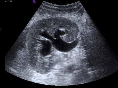
| Fig. 1 |
| Sonogram: Longitudinal image of the transplant kidney demonstrating hydronephrosis. |
|
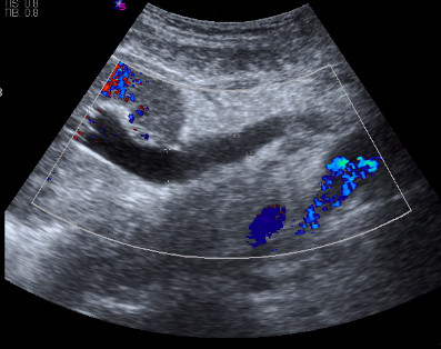 |
| Fig. 2 |
| Sonogram: Longitudinal image of the transplant ureter demonstrating ureteral dilatation. |
|
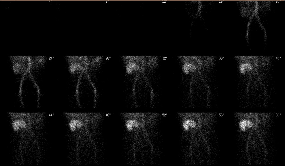 |
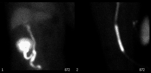
| Fig. 3 |
| Anterior pelvic radionuclide angiogram. |
|
|
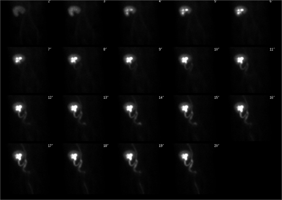 |
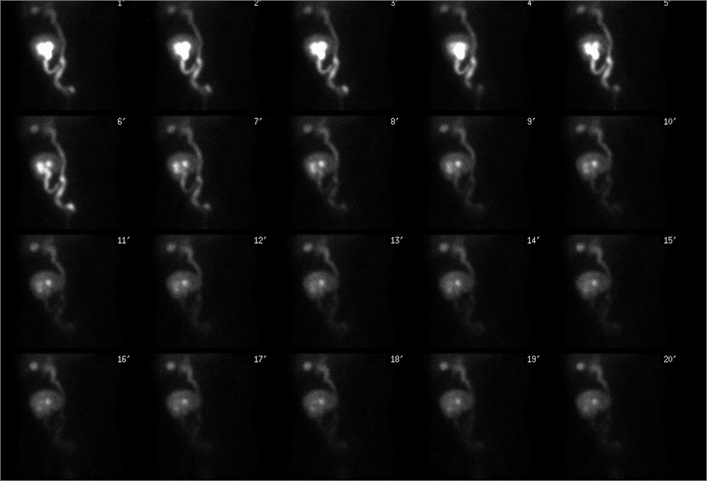
| Fig. 5 |
| Sequential anterior pelvic images of the transplant kidney through 20 minutes. |
|
 |
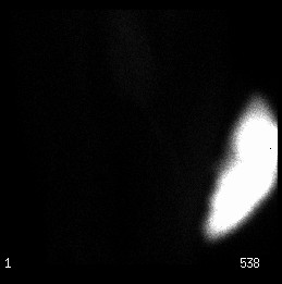
| Fig. 6 |
| Left: Anterior pelvic image. Right: Anterior image of the Foley catheter drainage bag (post void). |
|
 |
| Fig. 7 |
| Post-diuretic sequential anterior pelvic images through 20 minutes. |
|
 |
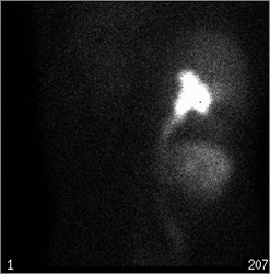
| Fig. 8 |
| Anterior image of the Foley catheter drainage bag (post diuretic). |
|
 |
| Fig. 9 |
| Posterior image of the native kidney: Activity is seen in collecting system as a result of reflux. Hepatic activity also is noted (a typical finding with Tc-99m MAG3. |
|