Return to Case List with Diagnoses or Case List as Unknowns

REFLUX VIA URETEROURETERIC ANASTAMOSIS
Authored By: tdeshm01 and Barry Siegel, Prof of Radiology.
Patient: 46 year old
History: 46-year-old man with vomiting and diarrhea.
Image Size:[small][as-submitted]

Fig. 1
Sonogram: Longitudinal image of the transplant kidney demonstrating hydronephrosis.

Fig. 2
Sonogram: Longitudinal image of the transplant ureter demonstrating ureteral dilatation.

Fig. 3
Anterior pelvic radionuclide angiogram.

Multimedia: 204432_4_submitted.avi

Fig. 5
Sequential anterior pelvic images of the transplant kidney through 20 minutes.

Fig. 6
Left: Anterior pelvic image. Right: Anterior image of the Foley catheter drainage bag (post void).

Fig. 7
Post-diuretic sequential anterior pelvic images through 20 minutes.

Fig. 8
Anterior image of the Foley catheter drainage bag (post diuretic).

Fig. 9
Posterior image of the native kidney: Activity is seen in collecting system as a result of reflux. Hepatic activity also is noted (a typical finding with Tc-99m MAG3.
Image Size:[small][as-submitted]

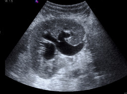
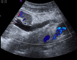
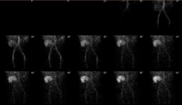
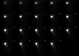
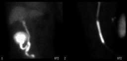
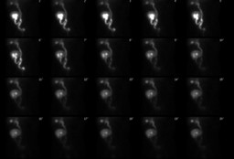
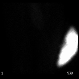
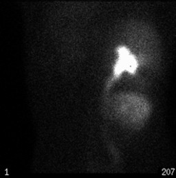
Findings:

RENAL SONOGRAM: Moderate hydronephrosis and hydroureter within the right transplant kidney.


RENAL SCINTIGRAPHY:

RADIOPHARMACEUTICAL: 7.5 mCi Tc-99m MAG 3 and 40 mg furosemide i.v.

FINDINGS: The anterior pelvic radionuclide angiogram demonstrates normal perfusion of the transplanted kidney in the right iliac fossa. The initial images demonstrate normal transplant size, morphology, and tracer accumulation. The sequential images show prompt uptake and excretion of the radiopharmaceutical by the transplant. The pelvicalyceal system of the transplant appears mildly dilated. Note also is made of reflux into the native right ureter via the uretoureteric anastomosis. The transplant ureter and the native ureter appear prominent. No abnormalities of the bladder are seen, and there is excretion of the tracer into the Foley catheter drainage bag. There is no evidence for urine extravasation or perirenal mass.

To evaluate for obstruction, the patient was given 40 mg furosemide via slow intravenous injection approximately 30 minutes after the start of the examination. Sequential images were obtained for an additional 20 minutes. There is prompt clearance of pelvicalyceal activity from the kidney after diuretic administration. A single static posterior image taken at the end of the examination additionally demonstrated activity within the native right renal collecting system, as a result of reflux.

Diagnosis:

1.Normal perfusion, morphology, and function of the renal transplant.

2. No evidence of obstruction of the transplant kidney.

3. Scintigraphic evidence of reflux of tracer into the right native ureter via the ureteroureteric anastomosis

General Discussion:

The patient underwent right renal transplant for end-stage renal disease 6 weeks previously. At surgery, the bladder was found to be extremely small and thick walled. Thus, it was decided to perform an end-to-side uretero-ureterostomy.The donor ureter was anastomosed to the native right ureter. A double-J ureteral stent was used to stent the anastomosis.

The patient presented with vomiting,diarrhea and an elevated serum creatinine. The ureteral stent was found to be malpositioned; hence, it was removed and a Foley catheter was placed.The renal scan was requested to evaluate the cause of transplant hydronephrosis and hydroureter seen on the subsequent sonogram.  

Specific Discussion:

Methods for reconstructing the urinary tract for renal transplantation include: 

1.  URETERONEOCYSTOSTOMY: The ureter is implanted into the bladder.  Advantages include the requirement for a very short length of ureter and maintenance of  the antireflux mechanism (by creating a tunnel). 

2.  URETEROURETEROSTOMY: The end of the donor ureter is anastomosed to the side of the recepient ureter.  This is preferred when the bladder wall is very thin or when the vascular attachments are such that a longer ureteral length would be required for bladder implantation. 

References: McDonald JC, Rohr MS, Frentz GD.  External ureteroneocystostomy and ureteroureterostomy in renal transplantationAnn Surg 1979; 190:663-7.
Comments:
No comments posted.
Additional Details:

Case Number: 204432Owner(s): tdeshm01 and Barry Siegel, Prof of RadiologyLast Updated: 02-07-2013
Anatomy: Genitourinary (GU)   Pathology: Iatrogenic
Modality: Nuc Med, USAccess Level: Readable by all users, writable by NucMed Certifiers
Keywords: rsnm

Case has been viewed 45 times.
Certified by Barry Siegel on 01-02-2010

The reader is fully responsible for confirming the accuracy of this content.
Text and images may be copyrighted by the case author or institution.