Return to Case List with Diagnoses or Case List as Unknowns

PROXIMAL PORTION OF THE JEJUNUM BLEEDING
Authored By: Keith Fischer and Xiaoni Hong.
Patient: 56 year old female
History:

56-year-old woman who presents with weight loss and bloody diarrhea.


Image Size:[small][as-submitted]

Fig. 1
GI bleeding Scintigraphic blood flow images reveal normal distribution of the radiopharmacetical.

Fig. 2
Sequential abdominal images were obtained through 60 minutes. There is a focus of increased activity that is seen in the left upper quadrant on the 5 minute image. The activity persists and travels anterograde into loops of small bowel during the remainder of the imaging period.
Image Size:[small][as-submitted]

Findings:

Following intravenous administration of Tc-99m labeled red cell, sequential abdominal images were obtained through 60 minutes. There is a focus of increased activity that is seen in the left upper quadrant on the 5 minute image. The activity persists and travels anterograde into loops of small bowel during the remainder of the imaging period. This is most likely in the proximal portion of the jejunum.


Diagnosis: Proximal portion of the jejunum bleeding
General Discussion:

Long history: 56-year-old woman who presents with weight loss and bloody diarrhea. She had recurrent intermittent lower gastrointestinal bleeding. The patient  is transferred from an outside hospital for decreased HCT. The HCT was 17% during the first few days of the admission and she required numerous tranfusions and two separate ICU stays during admission. Evaluate for bleeding source.


Radiopharmaceutical: 27.01 mCi Tc-99m in vitro labeled red cells i.v.

Angiography: No active extravasation or definite abnormal vascularity was identified on the mesenteric angiogram.


Follow up:  Small bowel enteroscopy shows an oozing linear jejunal ulcer with a visible vessel in the proximal jejunum.

Surgical Pathology Report: The jejunal biopsy revealed moderate villous blunting
and increased chronic inflammatory cells in the epithelium and lamina propria. 
Granulomas are absent.  These findings are not specific, but are consistent with
Crohn's enteritis.

Discussion: Bleeding from the small bowel is a rare cause of GI blood loss. Cancers, inflammatory bowel disease, and infection account for 20-25% of all small bowel bleeding, while AVMs account for the vast majority of bleeds. 
Determining the source of gastrointestinal bleeding that originates in the small bowel is one of the major diagnostic challenges facing gastroenterologists because the small bowel is long and hard to reach by endoscopy and therefore difficult to evaluate. 


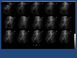
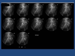
The GI bleeding scintigrahic study is to diagnose and localize the site of  bleeding. Over 80% of bleeding sites are detected during the initial imaging.  The activity persists and travels anterograde into loops of small bowel during the reemaider of the imaging period. This is most likely in the proximal portion of the jejunum.

References:

1.Gastroenterology; Volume 118, Issue 1, January 2000, Pages 201-221

2. Small bowel bleedong and capsule endoscopy. Website of the American College of Gastroenterology

3.The requisites Nuclear Medicine,third edition,page 369-371


Comments:
No comments posted.
Additional Details:

Case Number: 98003Owner(s): Keith Fischer and Xiaoni HongLast Updated: 02-07-2013
Anatomy: Gastrointestinal (GI)   Pathology: Vascular
Modality: Nuc MedAccess Level: Readable by all users, writable by NucMed Certifiers
Keywords: ginmACR: 70000.12170

Case has been viewed 33 times.
Certified by Keith Fischer on 11-02-2010

The reader is fully responsible for confirming the accuracy of this content.
Text and images may be copyrighted by the case author or institution.