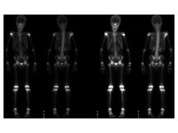
Delayed bone scintigraphy shows increased uptake seen within the right scapula.a second area of minimally increased uptake seen in the mid-shaft of the right humerus.
Fig. 2
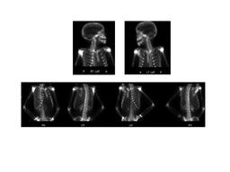
Delayed bone scintigraphy shows increased uptake seen within the right scapula.a second area of minimally increased uptake seen in the mid-shaft of the right humerus.
Fig. 3
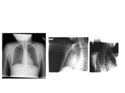
X-Ray of scapula, Chest reveals lytic lesions seen in the mid-diaphysis of the right humerus and the right scapula.
Fig. 4
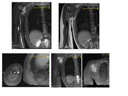
MRI reveals Large exophytic bony lesion emanating from the body of the right scapula, with large soft tissue component. An enhancing lesion is seen in the mid shaft of the right humerus.metastasis.
Case [View Case with Diagnosis]Owner(s): Xiaoni Hong and Akash SharmaLast Updated: 02-07-2013 Anatomy: Skeletal System Pathology: Other Access Level: Readable by all users, writable by NucMed Certifiers Keywords: bsnm
Case has been viewed 46 times. Certified by Akash Sharma on 10-29-2010
The reader is fully responsible for confirming the accuracy of this content. Text and images may be copyrighted by the case author or institution.