Return to Case List with Diagnoses or Case List as Unknowns

LANGERHANS' CELL HISTIOCYTOSIS
Authored By: Xiaoni Hong and Akash Sharma.
Patient: 8 year old female
History: 8-year-old girl with pain.
Image Size:[small][as-submitted]

Fig. 1
Delayed bone scintigraphy shows increased uptake seen within the right scapula.a second area of minimally increased uptake seen in the mid-shaft of the right humerus.

Fig. 2
Delayed bone scintigraphy shows increased uptake seen within the right scapula.a second area of minimally increased uptake seen in the mid-shaft of the right humerus.

Fig. 3
X-Ray of scapula, Chest reveals lytic lesions seen in the mid-diaphysis of the right humerus and the right scapula.

Fig. 4
MRI reveals Large exophytic bony lesion emanating from the body of the right scapula, with large soft tissue component. An enhancing lesion is seen in the mid shaft of the right humerus.metastasis.
Image Size:[small][as-submitted]

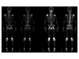
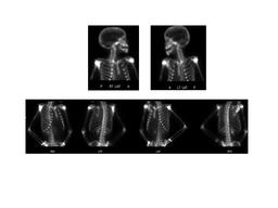
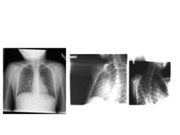
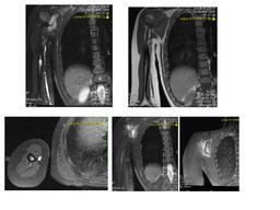
Findings:

Bone Scan: Delayed whole-body scintigrams were obtained.
There is increased uptake seen within the right scapula. This intensely increased uptake also demonstrates a central area with slightly less intense uptake in the remainder of this lesion. This may related to a history of recent biopsy in this location. There is a second area of minimally increased uptake seen in the mid-shaft of the right humerus,which corresponds to abnormality seen on recent MRI and plain radiographs.

X-Ray of scapula, Chest: 1. Lytic lesions in the mid-diaphysis of the right humerus and the right scapula, consistent with the patient's known diagnosis of eosinophilic granuloma without additional lytic lesions identified.

MRI: Large exophytic bony lesion emanating from the body of the right scapula, with large soft tissue component, concerning for Ewing sarcoma. An enhancing lesion is seen in the mid shaft of the right humerus.

DDx:

Primary tumor

   - Ewing’s  sarcoma

   - Osteosarcoma

   - Chondrosarcoma


Metastatic tumor


Langerhans cell histiocytosis


Infection


Fracture


Diagnosis: LANGERHANS' CELL HISTIOCYTOSIS
General Discussion:

Long history: 8-year-old girl with Langerhans' cell histiocytosis. The study is requested to evaluate extent of skeletal involvement. Comparison is made to a recent chest radiograph performed 11-8-05 and an MRI study performed on 11-4-05


Radiopharmaceutical: 7.6 mCi Tc-99m MDP i.v.

Follow up: BONE, RIGHT SCAPULA BIOPSY

     - LANGERHANS' CELL HISTIOCYTOSIS

BONE, RIGHT SCAPULAR MASS, LIMITED CURETTAGE

     - LANGERHANS' CELL

Specific Discussion:  Langerhans cell histiocytosis (LCH) is a group of idiopathic disorders characterized by the proliferation of specialized bone marrow–derived Langerhans cells (LCs) and mature eosinophils. The pathogenesis of LCH is unknown.LCH is a rare disease. The estimated annual incidence ranges from 0.5-5.4 cases per million persons per year. Approximately 1200 new cases per year are reported in the United States.
The prevalence of LCH seems to be higher among whites than other races.  The incidence of LCH is greater in males than in females, with a male-to-female ratio of 2:1. LCH affects patients from neonates to adults. The age at onset varies according to the variety of LCH.
  • Letterer-Siwe disease occurs predominantly in children younger than 2 years.
  • The chronic multifocal form, including Hand-Schüller-Christian syndrome, has a peak of onset in children aged 2-10 years.
  • Localized eosinophilic granuloma occurs mostly frequently in those aged 5-15 years.
  • Signs of LCH depend on the localization and the extent of the disease.
  • Imaging Studies:

    • Chest radiographs (posteroanterior and lateral)
      • LCH can present as a micronodular and interstitial infiltrate in the mid zone and base of the lung, with sparing of the costophrenic angles.
      • Older lesions show a honeycomb appearance.
      • Patients with radiographically demonstrated pulmonary involvement, in whom chemotherapy is being considered, require a biopsy of the lung preceded by bronchoalveolar lavage (BAL) to exclude opportunistic infections. If the BAL is diagnostic, the biopsy of the lung can be obviated.
    • Skeletal radiograph survey
      • Unifocal LCH presents as a single osteolytic lesion, usually affecting long or flat bones (in children, the calvaria and the femur; in adults, the ribs).
      • Multifocal LCH show osteolytic lesions involving the calvaria, the sella turcica, the mandible, the vertebrae, and/or the long bones of the upper extremities.
      • Although a radionuclide bone scan is suggested for establishing the extent of osseous involvement, the latter is not as sensitive as the skeletal radiograph survey in most patients.
    • CT scan or MRI of the hypothalamic-pituitary region may reveal abnormalities of these organs. In particular, magnetic resonance spectroscopy may be valuable in the early detection and evaluation of the neurodegenerative component.
References: emedicine:Langerhans cell histiocytosis (LCH)
Comments:
No comments posted.
Additional Details:

Case Number: 91792Owner(s): Xiaoni Hong and Akash SharmaLast Updated: 02-07-2013
Anatomy: Skeletal System   Pathology: Other
Access Level: Readable by all users, writable by NucMed Certifiers
Keywords: bsnm

Case has been viewed 46 times.
Certified by Akash Sharma on 10-29-2010

The reader is fully responsible for confirming the accuracy of this content.
Text and images may be copyrighted by the case author or institution.