back to index of exported cases

Unknown Case
Authored By: Garima Agrawal and Keith Fischer.
Patient: 54 year old female
History: 54 year old female with right breast cancer status post mastectomy and breast reconstruction in 2008. Recent chest CT demonstrated two sclerotic foci, one in the right sixth rib and another in the left seventh rib concerning for metastasis.  It was also suggested that healing fractures could have a similar appearance.
Image Size:[small][as-submitted]

Fig. 1
Recent CT chest study

Fig. 2
Whole body bone scintigraphy

Fig. 3
Spot images

Fig. 4
Whole body bone scintigraphy 4 years ago

Fig. 5
Radiograph of paranasal sinuses
Image Size:[small][as-submitted]

Comments:
No comments posted.
Additional Details:

Case [View Case with Diagnosis]Owner(s): Garima Agrawal and Keith FischerLast Updated: 02-07-2013
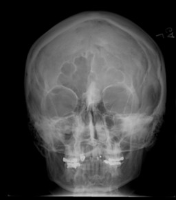
Anatomy: Face and Neck   Pathology: Benign Mass, Cyst
Modality: Conventional Radiograph, Nuc MedAccess Level: Readable by all users, writable by NucMed Certifiers
Keywords: bsnm, frontal, osteoma, scintigraphy

Case has been viewed 18 times.
Certified by Keith Fischer on 01-23-2013

The reader is fully responsible for confirming the accuracy of this content.
Text and images may be copyrighted by the case author or institution.