Return to Case List with Diagnoses or Case List as Unknowns

FRONTAL OSTEOMA ON BONE SCINTIGRAPHY
Authored By: Garima Agrawal and Keith Fischer.
Patient: 54 year old female
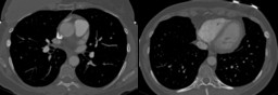
History: 54 year old female with right breast cancer status post mastectomy and breast reconstruction in 2008. Recent chest CT demonstrated two sclerotic foci, one in the right sixth rib and another in the left seventh rib concerning for metastasis.  It was also suggested that healing fractures could have a similar appearance.
Image Size:[small][as-submitted]

Fig. 1
Recent CT chest study

Fig. 2
Whole body bone scintigraphy

Fig. 3
Spot images

Fig. 4
Whole body bone scintigraphy 4 years ago

Fig. 5
Radiograph of paranasal sinuses
Image Size:[small][as-submitted]

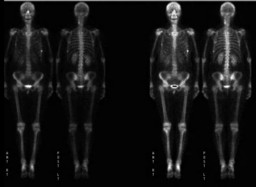
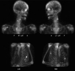
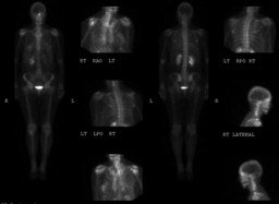
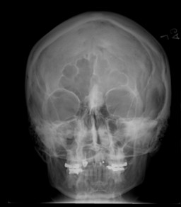
Findings:

Figure 1: Healing fractures of left 7th and right 6th rib.

Figure 2&3: There is focally increased activity at the anterolateral aspect of the left seventh and lateral aspect of the right sixth rib corresponding to  sclerotic lesions seen on CT. The scintigraphic pattern is more likely to represent fractures than metastases given there are no additional lesions elsewhere. There are no other new foci of increased activity. There is a stable focus of increased activity at the base of the left frontal sinus corresponding to a sclerotic lesion seen on prior radiograph of the paranasal sinus most consistent with an osteoma.

Figure 4: Prior bone scan showing similar findings of a focus of increased activity at the base of the left frontal sinus corresponding to a sclerotic lesion seen on radiograph of the paranasal sinus performed on the same day, most consistent with an osteoma.

Figure 5: Caldwell view of the paranasal sinuses at the time of prior bone scan demonstrates a well marginated lobulated 2.7 cm sclerotic density in the midline and the medial aspect of the left ethmoid sinus. Its appearance is consistent with an osteoma of the frontal bone.

DDx:

Frontal osteoma

Frontal intraasseous meningioma

Fibrous dysplasia

Chronic frontal sinusitis with inflammatory changes in the bone.

Diagnosis: Frontal osteoma and traumatic rib fractures.
General Discussion:

This patient had a focus of increased activity at the base of the left frontal sinus corresponding to a sclerotic lesion seen on prior radiograph of the paranasal sinus which remained stable for over 4 years most consistent with a diagnosis of frontal sinus osteoma.

Osteomas are usually asymptomatic and discovered incidentally in about 3% of the population undergoing paranasal sinus CT examination. These are most commonly found in the frontal sinuses in 80% of the cases followed by ethmoid air cells in about 15% and maxillary sinuses in about 15%. On plain radiograph these appear as well circumscribed dense lesions usually less than 2 cm in size. Bone scintigraphy often shows increased uptake in these lesions. These are usually subclassified as ivory osteoma (uniformly very dense) or mature osteoma (resembles 'normal' bone, including trabecular bone sometimes with visualization of marrow space). Frontal sinus osteomas with increased activity on bone scintigraphy have been shown to have potential for growth, while cold lesions tend to remain stable in size. Not very often these may cause obstruction of the sinus ostium leading to infection or mucocele formation. Very infrequently these may erode through the dura and result in CSF rhinorrhea or intracranial infection. These lesions are treated only if they are symptomatic or are growing in size. Endoscopic excision is the preferred treatment.

References:

http://radiopaedia.org/articles/paranasal-sinus-osteoma

Radionuclide bone scan in frontal sinus osteoma. Aust N Z J Surg. 1989 Feb;59(2):127-32.

Eur Arch Otorhinolaryngol. 2011 Apr;268(4):525-32. Epub 2010 Sep 17.
Comments:
No comments posted.
Additional Details:

Case Number: 389056Owner(s): Garima Agrawal and Keith FischerLast Updated: 02-07-2013
Anatomy: Face and Neck   Pathology: Benign Mass, Cyst
Modality: Conventional Radiograph, Nuc MedAccess Level: Readable by all users, writable by NucMed Certifiers
Keywords: bsnm, frontal, osteoma, scintigraphy

Case has been viewed 18 times.
Certified by Keith Fischer on 01-23-2013

The reader is fully responsible for confirming the accuracy of this content.
Text and images may be copyrighted by the case author or institution.