Return to Case List with Diagnoses or Case List as Unknowns

FAILURE OF FREE FLOW OF TRACER INTO THE PERITONEAL CAVITY
Authored By: gagraw01 and Barry Siegel, Prof of Radiology.
Patient: 39 year old male
History: 39-year-old man with well-differentiated mucinous adenocarcinoma of the appendix and pseudomyxoma peritonei, status post terminal ileum resection, appendectomy and partial colectomy in September 2009. An exploratory laparotomy, tumor debulking, peritoneal stripping, pancreatectomy, splenectomy and ileocolic resection was performed in July 2010 for residual mucinous adenocarcinoma. Intraperitoneal oxaliplatin therapy was planned.
Image Size:[small][as-submitted]

Fig. 1
Right peritoneal port injection

Fig. 2
left peritoneal port injection

Fig. 3
Transmission scan
Image Size:[small][as-submitted]

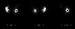
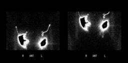
Findings: Loculated intraperitoneal tracer collections posterolaterally around the right and left peritoneal catheters, respectively. There is no free flow of the tracer from either of the catheters into the remainder of the peritoneal cavity.
DDx: Bilateral intraperitoneal loculations at catheter sites.
Diagnosis:

Bilateral intraperitoneal loculations at catheter sites.

General Discussion:

Peritoneal cavity scintigraphy involves intraperitoneal administration of Tc-99m sulfur colloid followed by promoting dispersion of the tracer through out the intraperitoneal cavity by rolling the patient from side to side and, thereafter, acquiring images in anterior and lateral projections and subsequently a transmission scan for better localization.  SPECT and/or SPECT/CT may also be helpful.

Peritoneal cavity scintigraphy is indicated for assessing direct communication between the peritoneal cavity and an extraperitoneal fluid collection (e.g., pleural effusion, hydrocele) and for evaluating the intraperitoneal distribution of tracer before intracavitary chemotherapy or radionuclide therapy of peritoneal malignancies in order to avoid excessive local chemotherapy concentrations or radiation doses in cases of loculations from intraperitoneal adhesions.

Specific Discussion:

Peritoneal mucinous carcinomatosis is a rare, malignant, intra-abdominal neoplasm that produces large amounts of mucin. Patients typically present with diffuse peritoneal disease. After surgical treatment, multiple locoregional recurrences are common [1].

Maximum cytoreductive surgery followed by consolidated intraperitoneal chemotherapy has emerged as a promising and potentially curative treatment option for patients with peritoneal dissemination of appendiceal mucinous tumors [2].

For these therapeutic approaches to be successful there must be free distribution of the therapeutic agent throughout the peritoneal cavity, maximizing contact of the agent with the tumor.

Single-photon emission computed tomography SPECT of the peritoneal cavity is a useful adjunct to planar imaging by adding spatial detail of anatomic areas impossible to resolve on planar images, and allowing for the visualization of the complexity of the peritoneal space [3].

References:

1.  Schomas DA, Miller RC, Donohue JH, Gill S, Thurmes PJ, Haddock MG, Quevedo JF, Gunderson LL. Lancet Oncol. 2006 Jan;7(1):69-76. Intraperitoneal treatment for peritoneal mucinous carcinomatosis of appendiceal origin after operative management: long-term follow-up of the Mayo Clinic experience. Ann Surg. 2009 Apr;249(4):588-95.

2.  Sugarbaker PH. New standard of care for appendiceal epithelial neoplasms and pseudomyxoma peritonei syndrome? Lancet Oncol. 2006 Jan;7(1):69-76.

3.  Wahl RL, Gyves J, Gross BH, Cochran M, Juni JE, Arnstein NB, Lahti D, Ackermann RJ. SPECT of the peritoneal cavity: method for delineating intraperitoneal fluid distribution. AJR Am J Roentgenol. 1989 Jun;152(6):1205-10.

Comments:
No comments posted.
Additional Details:

Case Number: 276236Owner(s): gagraw01 and Barry Siegel, Prof of RadiologyLast Updated: 02-07-2013
Anatomy: Gastrointestinal (GI)   Pathology: Neoplasm
Modality: Nuc MedAccess Level: Readable by all users, writable by NucMed Certifiers
Keywords: peritoneal, scintigraphy, mcnm

Case has been viewed 31 times.
Certified by Barry Siegel on 11-20-2010

The reader is fully responsible for confirming the accuracy of this content.
Text and images may be copyrighted by the case author or institution.