back to index of exported cases

Unknown Case
Authored By: Joanna Fair and Barry Siegel, Prof of Radiology.
Patient: 64 year old
History: Elevated 6-HIAA.
Image Size:[small][as-submitted]

Multimedia: 129392_1_submitted.avi
Maximum-intensity-projection (MIP) images of the processed SPECT data from an In-111 pentetreotide scan, obtained 24 hours post injection.

Fig. 2
Same reconstructed SPECT data, displayed as transaxial images.

Multimedia: 129392_3_submitted.avi
Raw data (projection images) from the same scan.

Multimedia: 129392_4_submitted.avi
Subset (few frames) of projection data showing the data collapse into a single point before disappearing.

Multimedia: 129392_5_submitted.avi
Repeat imaging of the same patient acquired 2 hours later. Maximum- intensity-projection (MIP) images are shown.
Image Size:[small][as-submitted]

Comments:
No comments posted.
Additional Details:

Case [View Case with Diagnosis]Owner(s): Joanna Fair and Barry Siegel, Prof of RadiologyLast Updated: 02-07-2013
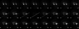
Anatomy: Gastrointestinal (GI)   Pathology: Other
Modality: Nuc MedAccess Level: Readable by all users, writable by NucMed Certifiers
Keywords: otnm, spect, artifact, octreotide, octreoscan, somatostatin receptorACR: 70000.93000

Case has been viewed 52 times.
Certified by Barry Siegel on 09-19-2009

The reader is fully responsible for confirming the accuracy of this content.
Text and images may be copyrighted by the case author or institution.